
Abstract
Introduction
Thymolipoma may be identified incidentally during a diagnostic screening for other medical problems and may grow large dimensions and manifest themselves clinically by signs of compression on adjacent structures such as shortness of breath, cough, palpitations, hypotension, tachypnea, the fullness of the neck veins and edema in the lower extremities [4].
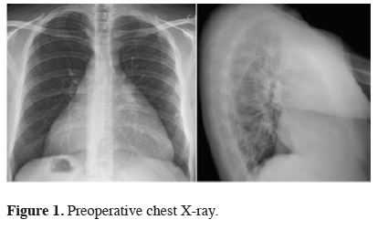
It can be seen at any age, but it is most common in children and in young ages. It is difficult to distinguish radiologically from other mediastinal masses, however, hyperplasic thymus and adipose tissue-like density may be determinative in contrast-enhanced computed tomography of the chest. We describe the case of a completely excised giant thymolipoma via uniportal video-assisted thoracoscopic surgery (VATS) which was detected incidentally on chest X-ray, with the review of the literature.
Case Presentation
)
 Click Here to Zoom |
Figure 1: Preoperative chest X-ray. |
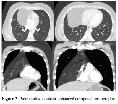
After being healed from the emergency situation, a contrast-enhanced computed tomography of chest (CCT) was taken under elective conditions. A massive soft tissue mass of fat density and giant size was observed, which completely filled the anterior mediastinum, extending from the right paracardiac area to the diaphragm, with clear borders with the mediastinal structures, not disrupting the contour of the mediastinal tissues (lesion dimension; 213x65 mm) (Figure 2).
)
 Click Here to Zoom |
Figure 2: Preoperative contrast-enhanced computed tomography. |
Although there was no additional disease in his medical history, when his anamnesis was deepened, it was found that there was shortness of breath and fatigue those had existed for a while and came with effort, were observed. Results of other clinical laboratory tests were normal. Biopsy and additional radiological examination were not performed in this case, because the patient was considered to have benign anterior mediastinal mass lesion and surgery was planned for complete excision. After the preoperative preparations were completed, the patient underwent left selective intubation, with a double-lumen endotracheal tube and the hemithorax was explored via uniportal VATS (3 cm incision) approach incision through the right fourth intercostal space on anterior axillary line. The mediastinal pleura was dissected and an encapsulated lesion of benign character was observed. After the thymic vein was clipped from the superior mediastinum, the left side mediastinal pleura was also opened (Figure 3).
)
 Click Here to Zoom |
Figure 3: Intraoperative anterior mediastinum and thymic vein image. |
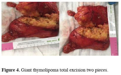
The capsule was preserved and the lesion was dissected from the pericardial and diaphragmatic surfaces with the help of electrocautery. At this point, the best effort was taken to preserve the phrenic nerve from an unintentional injury. The lesion could not be removed in one piece because of its giant size, was extracted with two separate endobags in two pieces, by dividing it into two in the endobag while maintaining the safe oncological principles (Figure 4).
)
 Click Here to Zoom |
Figure 4: Giant thymolipoma total excision two pieces. |
The incision was lengthened by 1 cm for lesion removal. Assistance was received from endoscopic and conventional surgical instruments that we constantly use in uniportal VATS application
Tube thoracostomy was performed from the same incision and no postoperative complication was observed in this case. On the second postoperative day, the chest tube was removed and on the third day, he was discharged after an uneventful course (Figure 5).
)
 Click Here to Zoom |
Figure 5: Uniportal VATS incision scar. |
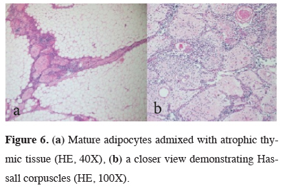
On macroscopic examination of the specimen, encapsulated two masses weighing totally 666 g, with the size of 16.5x11x3.5 cm and 13x10.5x3.5 cm were detected. The cut surface of the masses was solid and yellow-colored. Histopathologically, the tumor was reported to be thymolipoma. It was characterized by mature adipose tissue surrounded by a thin fibrous capsule and admixed with strands of atrophic thymic tissue containing Hassall corpuscles and calcification in some areas (Figure 6). The patient has been followed up for 10 months without any problem.
)
 Click Here to Zoom |
Figure 6: (a) Mature adipocytes admixed with atrophic thymic tissue (HE, 40X), (b) a closer view demonstrating Hassall corpuscles (HE, 100X). |
Written informed consent was obtained from the patient for publication of his data.
Discussion
The surgical approach preferred in mediastinal lesions growing huge dimensions is sternotomy and thoracotomy [2,10]. Thoracoscopic surgery has not been recommended due to the inability to remove these giant size lesions completely, the spread of tumor cells to the pleura and the risk of recurrence and VATS applicability has been reported only in small size lesions [10,11]. Uniportal VATS is a commonly used surgical approach in our clinic for mediastinal lesions. This surgery was completed with right uniportal VATS because it was considered as a benign and encapsulated lesion. Since the lesion was too large to be extracted with a single endobag, the giant lesion dissected from all mediastinal structures was divided into two parts in the endobag and completely extracted with two separate endobags.
Radiologically, lipoma, liposarcoma, teratoma, cardiomegaly, pericardial fat pad, pericardial effusion, pericardial tumors, congenital diaphragm hernias should be considered in the differential diagnosis. Making the correct diagnosis for thymolipoma by fine-needle aspiration cytology is not always possible, because the cytological material should contain both thymic and lipomatous components for differential diagnosis with other lipomatous tumors. In our opinion, it is not very satisfactory to perform needle biopsy to such type of lesions for diagnosis. In our case the differential diagnosis and the type of surgical approach were considered via CCT so that a preoperative biopsy was not indicated. Both definitive diagnosis and treatment were achieved with complete surgical resection.
Although the thymolipoma grows to huge dimensions, it may not show any symptoms and progress without symptoms. Mostly heavier than 500 g when diagnosed. In our case was asymptomatic and weighed 666 g.
As a conclusion, although thymolipoma is a rare benign lesion with no malignant features and no recurrence, it can grow to giant sizes and cause compression signs and symptoms. Therefore, complete excision should be performed with surgery for diagnosis and treatment. It should be kept in mind by experienced clinics that VATS is a good choice of approach, particularly in cases considered to be benign, even in the tumors grown to huge sizes.
Declaration of conflicting interests
The authors declared no conflicts of interest with respect to the authorship and/or publication of this article.
Funding
The authors received no financial support.