Current Thoracic Surgery 2017 , Vol 2 , Num 2
Atypical pulmonary hydatidosis lesions mimicking other lung lesions
Ülkü Yazıcı1,Serdar Özkan2,Özgür Ömer Yıldız3
1Private Thoracic Surgeon, Ankara, Turkey
2Department of Thoracic Surgery, Medova Hospital, Konya, Turkey
3Department of Thoracic Surgery, Yenimahalle State Hospital, Ankara, Turkey
DOI : 10.26663/cts.2017.0012

Summary

Background: Hydatid cyst disease, which is mostly caused by Echinococcus granulosus, may result in high morbidity rates particularly in endemic regions, and mortality depending on its size and on the organs where it is located. While the disease can be diagnosed radiologically because of typical symptoms, it becomes more difficult to distinguish it from lung malignancies as the cysts get complicated.

Materials and Methods: 15 cases of pulmonary hydatid cyst that underwent clinical and radiological examinations and surgery after being diagnosed as lung abscess and lung cancer between 2008 and 2014 were retrospectively reviewed.

Results: Radiological features of the 80% of the cases in thoracic CT were mimicking tumour in the form of thick walled cavitary lesion, hilar mass, cystic necrotic solid mass or solid mass; and 20% of them had radiological features mimicking abscess in the form of thin walled cavitary lesion. Lobectomy was performed for one case, wedge resection was performed for two cases and cystotomy-capitonnage was performed for 12 cases.

Conclusions: It should be kept in mind that pulmonary hydatid cyst clinically and radiologically may mimic lung cancer and lung abscess.

Introduction

Hydatid cyst disease, which could develop in almost all organs especially in liver and lung by forming cystic lesions has got high morbidity and mortality rates and it is a significant public health issue. While there could be symptoms depending on the location and the size of the intact cysts, the majority of the cases are asymptomatic. Pressure on neighboring organs or expectoration of the cyst due to rupture are among the main symptoms. Diagnosis is usually through lung graphics and computed tomography (CT). Radiological images of the cystic lesions are homogenous, round or oval and well demarcated. Atypical or complicated hydatid cysts in lungs could clinically and radiologically resemble other lung malignancies [1,2]. Similarity of complicated hydatid cyst lesions with other lung malignancies causes difficulties in diagnosis and planning the surgical treatment.

Methods

In this study, 15 cases of pulmonary hydatid cyst that underwent clinical and radiological examinations and surgery after being diagnosed with lung abscess and lung cancer between 2008 and 2014 in our thoracic surgery clinic were retrospectively reviewed. During the diagnostic workup, lung graphics, computed tomography and positron emission tomography (PET-CT) were performed. Transthoracic needle biopsy was performed in three cases. The cases included in this study are the ones with hydatid cysts whose clinical and radiological features are not consistent with the symptoms and radiology of hydatid cysts and the cases which mimic lung cancer or lung abscess. The cases evaluated as lung abscess after the radiological examination underwent antibiotherapy for 4 to 6 weeks, and in case of no significant change in radiological findings, surgical treatment was decided. Surgical procedures were planned depending on the localisation and the size of the lesion and different surgical procedures including cystotomy-capitonnage and lobectomy were performed. 12 of the cases were diagnosed with hydatid cyst in intraoperative stage, while three of them were diagnosed with hydatid cyst in the postoperative stage.

Results

The average age of the 15 cases included in this study was calculated as 49.3 (between 17 and 63). 8 of the cases are male and 7 of them are female. 33% of the cases (n=5) complained of cough, 27% of the cases (n=4) chest pain, 27% of the cases (n=4) hemoptysis and 13% (n=2) chills and fever. 60% of the lesions were located in the right lung. Radiological features of the 80% of the cases (n=12) in thoracic CT were mimicking tumour in the form of thick walled cavitary lesion, hilar mass, cystic necrotic solid mass or solid mass (Figures 1,2) and 20% of them (n=3) had radiological features mimicking abscess in the form of thin walled cavitary lesion (Figure 3). Characteristics of the cases enrolled in the study are located in Table
1.


Click Here to Zoom
Figure 1: Thorax CT showing hydatid cyst as a hilar mass.


Click Here to Zoom
Figure 2: Thorax CT showing hydatid cyst as a solid mass.


Click Here to Zoom
Figure 3: Thorax CT showing hydatid cyst as an abscess.

Table 1: Features of the cases

Positron emission tomography was performed for five cases out of 12, clinical and radiological findings of which mimic lung cancer. Fluorodesoxyglucose (FDG) retention in PET-CT was calculated between 2 and 7.8 (SUVmax). None of the cases was detected with cystic or mass lesion in addition to lung lesion. Bronchoscopy was performed for all cases in the preoperative stage. No pathological findings were found as a result of bronchoscopy. Transthoracic needle biopsy was performed for three cases, radiological features of which resembled to solid mass. In the histopathological evaluation of biopsy materials, no diagnostic results were obtained in relation to malignancy and hydatid cyst. In light of existing radiological findings, lobectomy was performed for one case, wedge resection was performed for two cases and cystotomy-capitonnage was performed for 12 cases. Pathological results of all cases were reported as hydatid cyst.

Discussion

The majority of hydatid cyst diseases are caused by the larval stage of E. Granulosus affecting mostly the liver and lungs, respectively. While the majority of the cases have solitary lesions, there are also cases having multiple lesions. In 60% of the cases, it is located in the right lung and in 20% of the cases the lesions are bilateral [3,4]. In all 15 cases included in this study, the lesions were unilateral and solitary and 60% of them were located in the right lung.

Lung hydatid cysts usually are not symptomatic. Symptoms start to appear after the size of the cyst reaches 5 cm and create pressure in the neighboring organs or in the case of ruptured and complicated the cystic lesion [5]. Depending on the size and the localisation of the cyst, non-specific symptoms including cough, hemoptysis and chest pain occur [6]. The most frequent symptoms in the majority of the cases were non-specific ones such as cough, whilst some cases had symptoms such as fever and shivers which could occur after the cyst rupture.

diagnosis of hydatid cyst, tests such as indirect hemagglutination or indirect fluorescent antibody can be applied [7]. However due to the lack of specific diagnosis in the preoperative stage of the cases according to the clinical and radiological findings, serological tests for the diagnosis of hydatid cyst could not be performed. Having said that, despite the positive serological tests for Echinococcus granulosus, there are reports arguing that serological tests, which identify large cell lung carcinoma, can give false positive results [8].

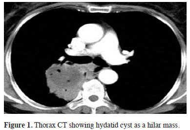
Since the clinical and laboratory findings are not specific in the diagnosis of hydatid cyst, radiological scan gains importance at the diagnosis stage [5]. Cysts that are not complicated are seen as well demarcated round or homogeneous masses in computed tomography and lung graphics. Larger cysts can result in atelectasis in lungs, mediastinal shift or pleural effusion depending on the level of the pressure on the neighbouring tissue. Lung graphics of the complicated cysts can show air-fluid level, water lily or meniscus signs. Ruptured cysts can appear as mass or abscess in computed tomography. Due to the fact that none of our cases had typical radiological findings for hydatid cyst and there were images mimicking abscess or tumour in direct graphics and computed tomography, the diagnostic and surgical approach to the cases showed differences. Five cases that were thought to be malignant were examined in PET-CT and the SUVmax values of these examinations [2-7,8] supported the malignancy diagnosis.

Due to the rise in glucose metabolism in the malignant pulmonary nodules, the FDG retention in the nodule also shows an increase. Lesions with FDG retention with SUVmax value higher than 2.5-3 in PET-CT are usually evaluated as malign [9]. However, as a result of rise in FDG retention due to infection and granulomatosis and inflammation, false positive results are not unexpected. The most frequent false positive results are seen in diseases including tuberculosis, sarcoidosis, aspergillus and parasitary diseases [10]. Local inflammation caused by cyst rupture in our cases resulted in positive PET-BT results for malignancy. Similar to our study, there are reports on breast carcinoma cases which were initially evaluated as pulmonary metastatic lesion in PET-CT and later was diagnosed as hydatid cyst during the surgical stage [11], or cases which underwent surgery due to the lesion which showed pulmonary hypermetabolic retention in PET-CT but later diagnosed as infected consolidated hydatid cyst during the pathological examination [12]. In another study conducted on this issue, it was reported that hydatid cyst disease can easily be confused with thoracic empyema, mediastinal mass, tuberculosis pleurisy and thoracic wall tumour [13].

As a conclusion it should be kept in mind that pulmonary hydatid cyst in endemic regions clinically and radiologically can mimic lung cancer and lung abscess.

Declaration of conflicting interests
The author declared no conflicts of interest with respect to the authorship and/or publication of this article.

Funding
The author received no financial support.

Reference

1) Kilinç O, Döskaya M, Sakar A, Yorgancioğlu A, Halilçolar H, Caner A, et al. Three atypical pulmonary hydatidosis lesions mimicking bronchial cancer from Turkey. New Microbiol 2009; 32: 229-33.

2) Ozkok A, Gul E, Okumus G, Yekeler E, Gulluoglu MG, Kiyan E, et al. Disseminated alveolar echinococcosis mimicking a metastatic malignancy. Intern Med 2008; 47: 1495-7.

3) Morar R, Feldman C. Pulmonary echinococcosis. Eur Respir J 2003; 21: 1069-77.

4) Deshmane JV. Multiple pulmonary hydatid cysts. Postgrad Med J 1967; 43: 774-8.

5) Sayek I. Hydatid Cyst Disease: Clinical Aspects. In: Altıntaş N, Tınar R, Coker A. editors. Echinococcosis. İzmir: Publications of Turkish Society of Hydatidology, 2004; 141-7.

6) Çobanoğlu U, Sayır F, Mergan D. Diagnostic dilemma: analysis of 11 cases of hydatid disease. Turkiye Parazitol Derg 2011; 35: 164-8.

7) Eşgin M, Aktaş M, Coşkun S. The investigation of antibody presence in the sera of patients with a suspicion of cystic echinococcosis by using indirect hemaglutination test (IHA). Turkiye Parazitol Derg 2007; 31: 283-7.

8) Singh N, Srinivas R, Bal A, Aggarwal AN. Lung carcinoma mimicking hydatid cyst: a case report and review of the literature. Med Oncol 2009; 26: 424-8.

9) Love C, Tomas MB, Tronco GG, Palestro CJ. FDG PET of infection and inflammation. Radiographics 2005; 25: 1357-68.

10) Kadam SG, Basu S, Joshi JM. Doughnut sign on FDG-PET scan in a ruptured lung hydatid cyst. Indian J Chest Dis Allied Sci 2012; 54: 185-7.

11) Kurt Y, Sücüllü I, Filiz AI, Urhan M, Akin ML. Pulmonary echinococcosis mimicking multiple lung metastasis of breast cancer: the role of fluoro-deoxy-glucose positron emission tomography. World J Surg Oncol 2008; 6: 7.

12) Rangarajan V, Dua S, Purandare NC, Shah S, Sharma AR. Pulmonary hydatid cyst detected on FDG PET-CT. Clin Nucl Med 2010; 35: 298-9.

13) Gürsoy S, Üçvet A, Türk F, Tözüm H, Erbaycu AE, Başok O. Diagnostic dilemma of hydatic disease: analysis of 16 patients. Turkiye Klinikleri J Med Sci 2009; 29: 457-63.