
Abstract
Introduction
Case Presentation
)
 Click Here to Zoom |
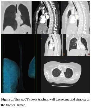
Figure 1: Thorax CT shows tracheal wall thickening and stenosis of the tracheal lumen. |
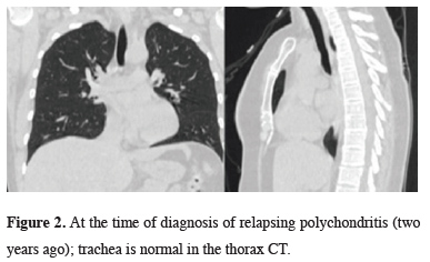
It was understood from the hospital records that the patient was diagnosed with RP due to swelling and edema in her ear 2 years ago. In the thorax tomography taken at this date, there was no thickening and stenosis in the wall of the trachea (Figure 2).
)
 Click Here to Zoom |
Figure 2: At the time of diagnosis of relapsing polychondritis (two years ago); trachea is normal in the thorax CT. |
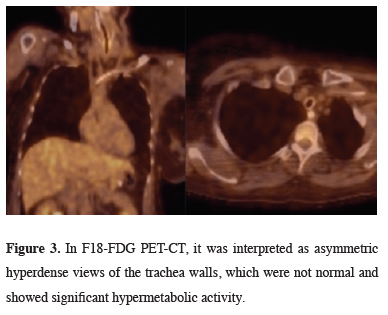
In PET/CT taken after thorax tomography, thick and asymmetric hyperdense views belonging to the tracheal walls, which did not show significant hypermetabolic activity, were detected (Figure 3).
)
 Click Here to Zoom |
Figure 3: In F18-FDG PET-CT, it was interpreted as asymmetric hyperdense views of the trachea walls, which were not normal and showed significant hypermetabolic activity. |
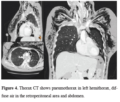
The patient was intervened with a rigid bronchoscope numbered 6.5. It was observed that the airways were hyperemic and edematous. No formation taking up endobronchial lesion was detected. However, during the maneuver with a rigid bronchoscope, the tracheal lumen was seen to collapse again. Tracheal stent implantation was planned for the patient whose extubation attempt was unsuccessful. During the stent application, the patient who developed CO2 retention stent attempt to the patient failed. The patient was intubated and returned to the anesthesia intensive care unit. Upon widespread development of subcutaneous emphysema, control thorax and abdominal CT were taken. There was pneumothorax in the left hemithorax, widespread air was detected in the retroperitoneal region and abdomen (Figure 4).
)
 Click Here to Zoom |
Figure 4: Thorax CT shows pneumothorax in left hemithorax, diffuse air in the retroperitoneal area and abdomen. |
Tube thoracostomy was performed to the left hemithorax. The patient underwent laparoscopy and the air in the abdomen was drained, but no perforation was detected. After re-bronchoscopy, mucosal laceration was detected in the left main bronchus in an area of approximately 0.5 cm. The chest drain was removed on the 6th day after the procedure after the follow-up of the patient, due to the expansion of the chest x-ray and no air and fluid drainage from the chest tube. The patient, whose medical treatment is continued in the intensive care unit; On the 11th day after laparoscopy, pseudomonas aeriginosa growth in bronchoalveolar lavage (BAL) culture. The patient died on the 28th day due to uncontrolled infection due to sepsis.
Written informed consent was obtained from the patient for publication of her data.
Discussion
In conclusion, relapsing polychondritis is a recurring inflammatory disorder of unknown etiology causing inflammatory reactions in the cartilaginous structures of the nose, ears, trachea, and the joints. Larynx, trachea, and bronchi are most frequently affected. Due to the rarity of the disease, there is no standardized treatment approach, and the treatment is guided by the clinical presentation and the severity of the disease [9]. The presented case was a patient who was diagnosed with relapsing polychondritis 2 years ago and developed tracheal involvement while under treatment, resulting in death. Detection of respiratory involvement at an early stage can significantly reduce mortality.
Declaration of conflicting interests
The authors declared no conflicts of interest with respect to the authorship and/or publication of this article.
Funding
The authors received no financial support.