
Abstract
Introduction
Here we aimed to present a rare and interesting chest wall metastasis case of lung cancer.
Case Presentation
)
 Click Here to Zoom |
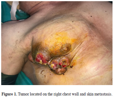
Figure 1: Tumor located on the right chest wall and skin metastasis. |
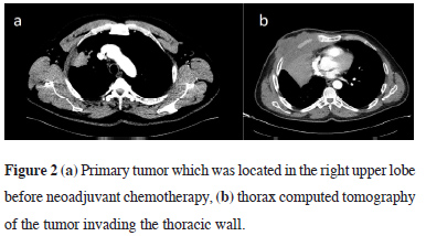
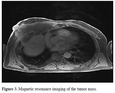
Thoracic CT and magnetic resonance imaging (MRI) were performed to evaluate the tumor size and borders (Figures 2, 3), and the patient was advised to consult with the plastic surgery clinic. Tumor resection and chest wall reconstruction were planned.
)
)
 Click Here to Zoom |
Figure 2: (a) Primary tumor which was located in the right upper lobe before neoadjuvant chemotherapy, (b) thorax computed tomography of the tumor invading the thoracic wall. |
 Click Here to Zoom |
Figure 3: Magnetic resonance imaging of the tumor mass. |
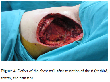
During surgery, the tumor, with the anterior portion of the right third, fourth, and fifth ribs (Figure 4), was resected.
)
 Click Here to Zoom |
Figure 4: Defect of the chest wall after resection of the right third, fourth, and fifth ribs. |
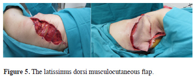
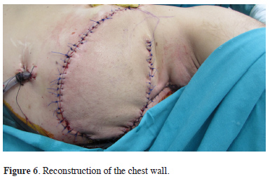
Tumor invasion into the lung parenchyma was not found. A latissimus dorsi musculocutaneous flap was used for reconstruction (Figures 5, 6).
)
)
 Click Here to Zoom |
Figure 5: The latissimus dorsi musculocutaneous flap. |
 Click Here to Zoom |
Figure 6: Reconstruction of the chest wall. |
In the lateral decubitus position, the latissimus dorsi muscle borders are marked and the incisions are planned according to the defect size. The adequate muscle is identified according to the deepness of the defect. Then the pedicle dissection is performed from the inferior angle of the scapula to the inferior. After that, the medial portion and the inferior portion of the flap were dissected, respectively. The flap was tunneled subcutaneously and adapted to the defect area. The patient’s pathology indicated lung epidermoid cancer metastasis. Postoperatively, the patient was followed up for 1 month.
Written informed consent was obtained from the patient for publication of his data.
Discussion
Evaluation of the tumor infiltrating the soft tissues or bones of the chest wall and preparation for complete chest wall resection are necessary to perform radical surgery. Invasion of the thoracic wall region can be evaluated using CT, bone scan, and MRI [3].
Lung cancer invasion of the chest wall is not a common occurrence, and clinical exploration is necessary for approximately 5% of patients who have undergone resection for lung cancer [4]. When complete resection is attempted, surgeons should use anatomical knowledge to choose the optimum route of access and increase the feasibility of radical resection because of the risk of recurrence. Complete resection is defined as a pathological demonstration of negative tissue margins via frozen sections. In the present case, the resection margin was intraoperatively confirmed using frozen sections for complete resection. The patient’s positive cytology findings indicated incomplete resection. Gross residual disease after attempted resection is also classified as palliative resection. If chest wall invasion is preoperatively suspected, chest wall resection including the affected rib should be scheduled.
Resection completeness, nodal status, invasion depth, tumor size, and adjuvant chemotherapy are the prognostic factors for the long-term survival of NSCLC patients with chest wall invasion.
A statistically significant difference in terms of survival rate has been reported among patients who underwent complete resection as compared to the rate among those who received incomplete resection.
The first three ribs are well covered by pectoral muscles and do not need a replacement, primary sutures are adequate. After resection of anterior ribs or more than three ribs to need grafts or free flaps. If you planned to resect ribs, which are located in the backside of the scapula or not enlarged than three ribs, reconstruction is not performed in principle. When necessary, reconstruction of the deficit of the chest wall without synthetic materials should be attempted as soon as possible. In the present case, a latissimus dorsi musculocutaneous flap was created through plastic surgery. Complications were not encountered due to the strength and stability of the mediastinum; neither a graft nor methyl methacrylate was necessary. Successful repair was confirmed by the free flap filling the thorax. Because the lung resection was previously performed, the intercostal spaces were narrowed on the same side and the muscle flap was sufficient for reconstruction. Besides, the mediastinum was stable and fixed due to radiotherapy, so mediastinal flutter and the flail chest were not expected.
The latissimus dorsi muscle obtains its blood supply from the thoracodorsal artery via the subscapular artery system. Thus, this long pedicle elevates this largest flap for the chest wall defects. The upper edge of this flap is the inferior angle of the scapula; when one is dissecting from superior to inferior, it is important to not dissect the serratus anterior muscle fascia [5]. The serratus muscle fascia should be left intact.
The latissimus dorsi flap allows for the inclusion of the required amount of skin and muscle. The latissimus dorsi musculocutaneous flap is a versatile flap for chest wall reconstruction [6].
In conclusion, pulmonary malignancies may be addressed by a wide spectrum of treatment approaches. Treatment options can be selected according to tumor localization. Chest wall metastasis is a very rare clinical condition requiring reconstruction after tumor resection.
Declaration of conflicting interests The authors declared no conflicts of interest with respect to the authorship and/or publication of this article.
Funding
The authors received no financial support.
Acknowledgement
We would like to offer special thanks to deceased Assoc. Prof. Dr. Murat Oncel, although no longer with us, for his support and efforts.