
Abstract
Materials and Methods: Patients who were admitted to our outpatient clinic or who were referred by other clinics for foreign body aspiration between January 2012 and January 2017 were retrospectively analyzed. Foreign objects such as coins, food pieces, toy pieces, and needles, which were routinely encountered, were excluded from the study. Gender, age, complaints, radiological findings, and surgical interventions were recorded.
Results: There were five cases (four males, one female) under the age of 18 and three male cases above the age of 18. The average age was 21.18 ± 28.93. There was one case with laryngectomy. In six cases, the foreign body was removed by rigid bronchoscopy. Lung resection was performed in three cases (one lobectomy and one wedge resection). The foreign bodies were a pen tip, a piece of stone, a rivet from jeans, the LED of a remote control, a latch spring, a piece of a spoon, a wood piece, and a piece of a dental filling.
Conclusions: Foreign body aspirations are among the most significant causes of morbidity and mortality, especially in the child age group. Diagnosis is possible through anamnesis, physical examination, and evaluation of radiological findings together. In the event of clinical suspicion, foreign body aspirations should be kept in mind regardless of age group.
Introduction
This study aims to examine interesting foreign bodies in terms of diagnosis and bronchoscopic removal methods, to emphasize that any foreign body can be aspirated in the child age group, and to present interesting foreign body aspirations encountered in adulthood.
Materials and Methods
Patient interventions included anamnesis, physical examination, and radiological examinations. Posteroanterior chest radiographs and computed thorax tomography were used as radiological examinations. In the presence of an object that was not detected radiologically in pediatric patients but that was suspected in anamnesis and physical examination, intervention was performed after families were informed about the relevant risks. All pediatric patients underwent rigid bronchoscopy (Karl Storz™, Germany) under general anesthesia. In patients presenting with intrathoracic lesions, thorax tomography was performed first. Surgery was performed in accordance with lung pathology.
Results
)
)
)
 Click Here to Zoom |
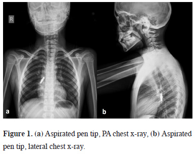
Figure 1: (a) Aspirated pen tip, PA chest x-ray, (b) Aspirated pen tip, lateral chest x-ray. |
 Click Here to Zoom |
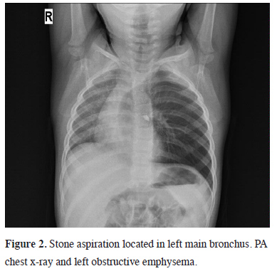
Figure 2: Stone aspiration located in left main bronchus. PA chest x-ray and left obstructive emphysema. |
 Click Here to Zoom |
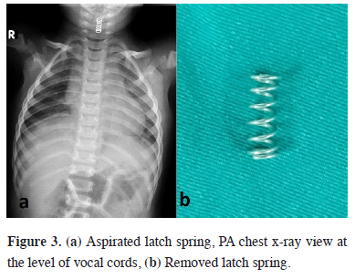
Figure 3: (a) Aspirated latch spring, PA chest x-ray view at the level of vocal cords, (b) Removed latch spring. |
A foreign body was observed in the right lower lobe of one case with a pulmonary complication, and in more distal of the airway in the left lower lobe of one case (Table 1).
)
Table 1: Characteristics of the patients.
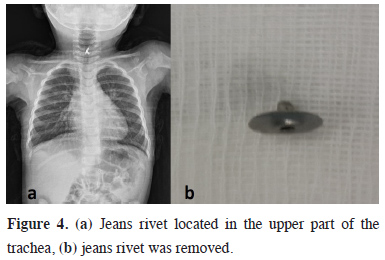
In only three of the cases, the anamnesis finding was a sudden cough; one adult patient admitted to the hospital after noticing aspiration of foreign body. The most common physical examination finding was ronchus. Fever was detected in one case. Although a foreign body history was taken in four cases, the physical examination findings were normal. In five of the cases, foreign bodies were detected by direct radiography due to their metal content (Figure 4).
)
 Click Here to Zoom |
Figure 4: (a) Jeans rivet located in the upper part of the trachea, (b) jeans rivet was removed. |
Pulmonary consolidation and fibroatelectatic infiltrations were detected in one of two patients who applied with pulmonary complications, while indirect radiological findings were detected in one case. Pneumonia was detected in one patient. A foreign body was detected by bronchoscopy in six of the cases who arrived in the early acute period. In five of all cases in which tracheobronchial foreign bodies detected, the foreign bodies were removed under general anesthesia, while one was removed under local anesthesia by rigid bronchoscopy. Bronchoscopy was performed in five cases within the first eight hours. One of the cases with chronic foreign body aspiration was detected after six years. Left lower lobectomy with thoracotomy was performed in one patient who was admitted with pulmonary complications, while video-assisted thoracoscopic wedge resection was performed in one patient.
Surgical Manipulations
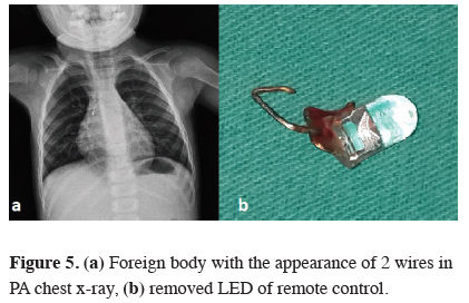
None of the patients developed acute respiratory failure. In six cases, foreign bodies were removed by bronchoscopy no later than 16 hours after aspiration. There was not any patient who underwent bronchoscopy developed pneumothorax in the postoperative period. Chest radiography was performed in each patient in the early postoperative period and 24 hours later. In cases in which patients applied with tracheal foreign body aspiration, and in which bronchoscopy was immediately performed, the foreign body was pushed to the right main bronchus, and the oxygen saturation was increased by ventilating the left lung. In one case, LED lamp wires were first passed through the bronchial orifice and then straightened and removed (Figure 5).
)
 Click Here to Zoom |
Figure 5: (a) Foreign body with the appearance of 2 wires in PA chest x-ray, (b) removed LED of remote control. |
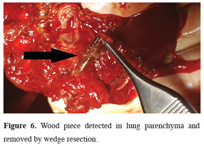
In delayed cases, although a bronchoscopy was performed for the etiology of recurrent infection and bronchiectasis, no endobronchial lesion was observed, and a foreign body was detected in the resection material. In one case, after the consolidation in the right lower lobe was removed by wedge resection, a hard body was detected during palpation of the parenchyma and a piece of wood was seen endobronchially (Figure 6, Table 2).
)
)
 Click Here to Zoom |
Figure 6: Wood piece detected in lung parenchyma and removed by wedge resection. |
Table 2: The findings of physical examination, the time of diagnosis and the treatment.
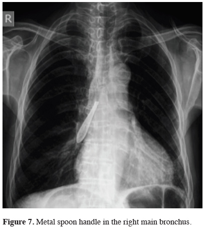
In one case who had permanent tracheostomy due to laryngectomy, the spoon handle that was aspirated during secretion cleaning was removed from the tracheostomy by bronchoscopy under local anesthesia (Figure 7).
)
 Click Here to Zoom |
Figure 7: Metal spoon handle in the right main bronchus. |
Discussion
Patients often present with acute respiratory distress and cough. Cough is caused by movement, irritation, and edema of the foreign body in the bronchus. After the foreign body has settled in the bronchi, the cough decreases and, depending on its localization, dyspnea often occurs [4-8].
Radiological evaluation should be performed in all cases in which a tracheobronchial foreign body is suspected; the diagnosis should be ruled out in individuals with normal radiology [9]. The nature of the aspirated foreign body affects the clinic. Tracheobronchial foreign bodies are often localized to the right bronchial system due to the anatomical angle. The right main bronchus is a continuation of the trachea, and is shorter and wider than the left. The dimensions of the angles in children are close to those in adults. In this study, four of the foreign bodies were seen in the right main bronchial system and two in the left bronchial system. No foreign body was detected in the carina. An interesting situation in another case was the pediatric patient who had emphysema on the right due to near right total bronchial obliteration, but who was initially diagnosed with asthma and was followed clinically for one year under this diagnosis. These rare occurrences show the importance of anamnesis and radiology in patients with a suspected foreign body.
Rigid bronchoscopy is a preferred method in children [5,6]. Thanks to its wide canal structure and lumen, the lung is easily ventilated by the anesthetist and bronchoscope also allows access to the foreign body [10]. Research suggests using steroids before and after bronchoscopy to reduce the incidence of postoperative subglottic edema that will require urgent tracheostomy [5-11]. For this reason, steroid treatment was started before bronchoscopy and continued for 24 hours at a dose of 1 mg/kg. During foreign body removal in foreign body aspiration cases, larynx edema, pneumomediastinum, pneumothorax, bleeding, and cardiac arrest can be seen [10-13].
Foreign body aspirations are rarely seen in adult patients. High-risk cases include the elderly, alcohol addicts, those with convulsions, users of sedative or hypnotic drugs, and people with neuromuscular disease, mental retardation, or dental prosthesis interventions [14,15]. In this study, one of the adult patients aspirated the foreign body through the permanent tracheostomy.
Late-diagnosed foreign body aspirations can be clinically present with lung and pleural complications. Complications can vary due to the localization of the foreign body, its type, and the time until diagnosis. Prolonged fever, hemoptysis, chronic cough, recurrent lung infections, bronchiectasis, bronchial stenosis, and lung abscess are some of the possible complications [16]. When irreversible conditions such as bronchiectasis or pulmonary abscess occur, lung resection is required [17,18]. Generally, lower lobes are affected and postoperative histopathological results show a foreign body in the lobar bronchus [19].
In conclusion, tracheobronchial foreign body aspirations are important causes of morbidity and mortality in childhood. Cases that are not noticed in the early period may present later with lung abscess, bronchiectasis, or bronchopneumonia. It should also be kept in mind that clinicians can encounter foreign bodies that are composed of both metallic and plastic components, or unusual foreign bodies that present with pneumonia, which require long diagnosis and treatment periods. Anamnesis and physical examination findings correlated with anamnesis are more important than the other examinations in pediatric populations. We recommend that endoscopic examinations be performed in every suspicious case and that if an unusual foreign body is detected, surgeons should use novel body removal methods and maneuvers besides conventional foreign body removal techniques before switching to the thoracotomy.
Declaration of conflicting interests
The authors declared no conflicts of interest with respect to the authorship and/or publication of this article.
Funding
The authors received no financial support for the research and/or authorship of this article.