
Summary
Introduction
The case was reported to avoid misdiagnosis and unnecessary surgery, due to the unusual location of EMH, by mimicking solid neoplasm in the posterior mediastinum as an unusual location. Besides, it was reported to raise awareness of hematological neoplasms that manifest themselves with anemia and form mass lesions.
Case Presentation
)
 Click Here to Zoom |
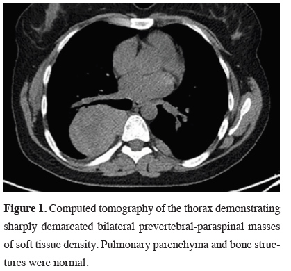
Figure 1: Computed tomography of the thorax demonstrating sharply demarcated bilateral prevertebral-paraspinal masses of soft tissue density. Pulmonary parenchyma and bone structures were normal. |
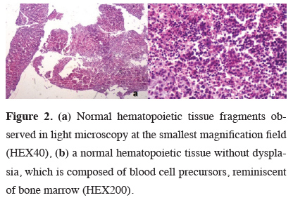
A tru-cut biopsy was performed from the mass that was located in the posterior mediastinum. Microscopically, the composition of cells were lymphocytes, erythrocytes, plasma cells, megakaryocytes and histiocytes (Figures 2a and b) where the structure showed similarity to bone marrow.
)
 Click Here to Zoom |
Figure 2: (a) Normal hematopoietic tissue fragments observed in light microscopy at the smallest magnification field (HEX40), (b) a normal hematopoietic tissue without dysplasia, which is composed of blood cell precursors, reminiscent of bone marrow (HEX200). |
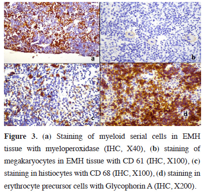
Immunohistochemical stains showed that some lymphocytes were focally stained with CD3, the other lymphocytes were stained with CD20. Megakaryocytes were positive with CD61 (Figure 3a), myelocytes were positive with MPO (Figure 3b). Erythrocyte precursor cells were found to be positive with GLCO A (Figure 3c) and monocytes were positive with CD68 (Figure 3d).
)
 Click Here to Zoom |
Figure 3: (a) Staining of myeloid serial cells in EMH tissue with myeloperoxidase (IHC, X40), (b) staining of megakaryocytes in EMH tissue with CD 61 (IHC, X100), (c) staining in histiocytes with CD 68 (IHC, X100), (d) staining in erythrocyte precursor cells with Glycophorin A (IHC, X200). |
The cells showed negative immunoreactivity with pancytokeratin, CD34 and CD117. In light of the whole clinical and histological features, this case was diagnosed as extramedullary hematopoiesis. With the final diagnosis, the patient referred to hematology clinic and blood transfusions with symptomatic management were applied. In her 10-months follow up period the whole process was uneventful.
Written informed consent was obtained from the patient for publication of her data.
Discussion
The causes of EMH may be pathological or physiological. There are wide varieties of pathologic conditions in which disturbance of the normal blood-forming marrow may be associated with the formation of extramedullary hematopoietic tissue. Common causes of EMH include spherocytic anemia, erythroblastosis of newborn, thalassemia, pernicious anemia in the period of relapse, macrocytic anemia of hepatic origin, carcinomas with bone marrow invasion, Hodgkin’s disease, lymphoma, leukemia, osteosclerosis and myeloproliferative syndrome [1-8].
The mechanism of origin of these extramedullary foci is unknown, but there are several theories like embolization, direct extension, myelostimulation or redirected differentiation, which explain the pathogenesis of EMH, but none of them is widely acceptable [15]. EMH is a physiologic compensatory mechanism that is usually associated with hematologic disorders as in our case and most of the other reported cases. EMH cases without hematological disorder have also been described [10].
Symptoms related to mass formation seen in EMH cases are related to the location of the lesion [2,10]. Patients may sometimes be asymptomatic [2,11]. Although the masses those are locating in the posterior mediastinum usually cause no symptoms, the ones with large dimensions might cause respiratory problems related to compression and neurologic deficits related to the level of involvement. The patients have complaints due to spontaneous pneumothorax, hemithorax and rapidly fatal respiratory failure [10,11,15]. Our case also had severe respiratory distress. Also, anemia was found in the patient who had complaints of weakness and fatigue.
It was determined that the case, which was immediately biopsied with the concern of the possibility of advanced neoplastic lesions, was a case of EMH secondary to anemia, forming a solid mass. A population of cells resembling bone marrow was observed in routine preparations obtained from the biopsy. Mesenchymal originated mediastinal neoplasms, especially lymphomas, were also included in the differential diagnosis. However, those were excluded by the presence of precursor cells, same as in the bone marrow, which constitutes blood cells examined in light microscopy. Possible lymphoreticular tissue neoplasms were also excluded from the differential diagnosis due to the absence of features such as atypia, pleomorphism, and disorganization, and determination of cell origin by additional immunohistochemical examinations.
Treatment of EMH diagnosed solid masses, can be variable depending on the patient and the disease. These methods are the use of blood transfusion, radiotherapy, surgical decompression, hydroxyurea and JAK2 inhibitors. JAK2 inhibitors and TGF-β ligand traps that improve the erythropoiesis process are being tested in patients with thalassemia and EMH who do not respond to treatment, and the results look promising. In the absence of severe symptoms and complications, EMH treatment may not be required [2]. Our patient whose symptoms started to regress with blood transfusion treatment is followed up by the clinic.
In conclusion, EMH is a rare pathology, but can be serious with mortal complications and also can be confused clinically and radiologically with other benign or malignant lesions in the posterior mediastinum. It should be kept in mind in the differential diagnosis of resistant anemia cases with radiologically determined mass. Clinician-radiologist-pathologist collaboration is required to avoid misdiagnosis.
Declaration of conflicting interests
The authors declared no conflicts of interest with respect to the authorship and/or publication of this article.
Funding
The authors received no financial support.