
Abstract
Introduction
Case Presentation
)
 Click Here to Zoom |
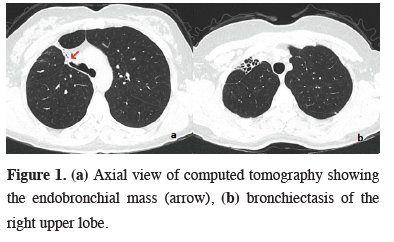
Figure 1: (a) Axial view of computed tomography showing the endobronchial mass (arrow), (b) bronchiectasis of the right upper lobe. |
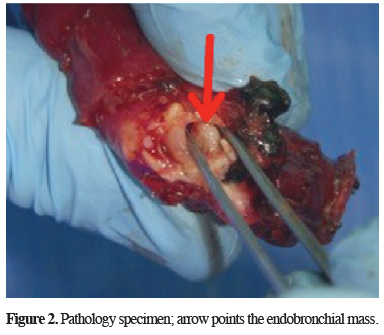
A fluorodeoxyglucose positron emission tomography (F-18 FDG PET/CT) was also administered at that hospital and it showed a minimally elevated maximum standard uptake volume (SUVmax) as 1.9. Bronchoscopy was performed in our hospital and the endobronchial mass was visualized as a smooth surfaced polypoid body at the entrance of posterior segmental bronchus of right upper lobe, which narrowed the bronchus of right upper lobe. A bronchoscopic biopsy was performed and its pathological examination reported superficial bronchial mucosa, indicating that the biopsy was taken superficially. We performed a video-thoracoscopic right upper lobectomy, since the video-thoracoscopic exploration revealed a totally shrunk right upper lobe and thinned out vasculature of the same lobe in accordance with preoperative CT scan. Another side note of the operation is that the lower and middle lobes were expanded compensatory to fill right hemithorax. An intraoperative frozen section was performed after the lobectomy and it reported the bronchial margins negative and also the endobronchial nodule was in benign nature (Figure 2).
)
 Click Here to Zoom |
Figure 2: Pathology specimen; arrow points the endobronchial mass. |
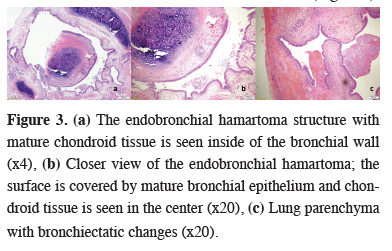
Histopathological examination of the specimen revealed the nodule as endobronchial hamartoma (Figure 3).
)
 Click Here to Zoom |
Figure 3: (a) The endobronchial hamartoma structure with mature chondroid tissue is seen inside of the bronchial wall (x4), (b) Closer view of the endobronchial hamartoma; the surface is covered by mature bronchial epithelium and chondroid tissue is seen in the center (x20), (c) Lung parenchyma with bronchiectatic changes (x20). |
Written informed consent was obtained from the patient for publication of her data.
Discussion
There are various cases in the literature which represented with recurrent pneumonia, persistent cough and even atrial fibrillation along with countless asymptomatic cases [2,4,5]. Some of these cases were reported to be with local bronchiectasis but there is no lobar bronchiectasis in the literature caused by an endobronchial tumor. Due to its nature, lobar bronchiectasis tends to reveal itself as recurrent pneumonia and frequent hospitalization and/or medication history [6]. In spite of our patient have had lobar bronchiectasis, there was no recurrent hospitalization or history of recurrent pneumonia. Nonetheless our patient showed persistent cough and mild hemoptysis, these symptoms does not necessarily indicate bronchiectasis, let alone lobar bronchiectasis.
Hamartomas, including endobronchial ones, incline to stay below-radar in F-18 FDG PET/CT [7,8]. Initial examination and evaluation of all findings, including CT scan and SUVmax value, directed the route towards low activity nodules of the lung: Typical carcinoid, pulmonary adenoma, bronchioloalveolar carcinoma and of course, endobronchial hamartoma [9-11]. There are many options in terms of treatment for endobronchial lesions. Bronchoscopic treatment may be the first one to think as a safe choice: diode laser or argon plasma coagulation and cryotherapy [12]. We have steered for an anatomical resection after a non-diagnostic bronchoscopic biopsy attempt, due to the localization of the mass and bronchiectatic appearance of the right upper lobe in preoperative CT scan. A bronchotomy also comes to mind, but the localization of the mass prevented the idea and also an anatomical resection was already inevitable due to bronchiectasis.
In conclusion endobronchial tumors may cause a variety of symptoms and problems. It is important to detect such masses in early stages, before the irreparable damage takes place. Bronchiectasis, as an irreversible damage entity of the lung, may be the outcome of endobronchial tumors. Our case showed that endobronchial hamartoma caused a total lobar bronchiectasis and is the sole case in the English literature to our knowledge.
Declaration of conflicting interests
The authors declared no conflicts of interest with respect to the authorship and/or publication of this article.
Funding
The authors received no financial support.