
Abstract
Introduction
Aberrant right subclavian artery (ARSA) is one of the rare reasons for dysphagia. In 80% of the cases it runs posterior to the esophagus to connect to aorta distal to the left subclavian artery and may cause catastrophic complications if unrecognized before surgical interventions in close proximity [4]. The recurrent laryngeal nerve also has an abnormal course in cases of aberrant right subclavian artery, branching from vagus, entering directly to larynx.
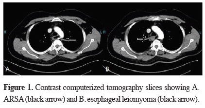
In this article, we report the coincidence of an esophageal leiomyoma and an ARSA in a patient who presented to our clinic with dysphagia.
Case Presentation
)
)
 Click Here to Zoom |
Figure 1: Contrast computerized tomography slices showing A. ARSA (black arrow) and B. esophageal leiomyoma (black arrow). |
 Click Here to Zoom |
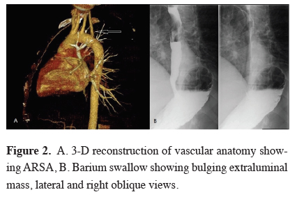
Figure 2: A. 3-D reconstruction of vascular anatomy showing ARSA, B. Barium swallow showing bulging extraluminal mass, lateral and right oblique views. |
The patient underwent a bi-portal (4th and 6th intercostal space, anterior axillary line) VATS enucleation of the leiomyoma in left lateral decubitus position. Dividing the azygous vein and a 3 cm myotomy was required to gain necessary exposure for safe removal of the tumor. The myotomy site was on the right lateral wall of the esophagus for careful avoidance of the ARSA. The myotomy was closed with three 2-0 absorbable sutures. The patient had an uneventful intra- and postoperative course and was discharged on 3rd postoperative day. He was maintained on a soft diet for 2 weeks and can consume a normal diet on 1st month of follow up, without any symptoms.
Discussion
In cases of mucosal rupture during stripping of the tumor, simple suturation is usually enough for leak control. In our case, we did not encounter any mucosal rupture, but placed sutures anyway, with the rationale of restoring the anatomy and the blood supply.
Endoscopic ultrasound is fairly specific for diagnosis of esophageal leiomyoma and is also confirmed by pathology results in larger case series. Biopsy is also proven to be safe in cases of submucosal tumors, but has little value for changing the clinical decision making if the resection is to be done for symptomatic purposes (dysphagia) [6,7].
VATS enucleation of esophageal leiomyoma has been reported in large case series and results have been more favorable than thoracotomy [5]. In our case, VATS approach did not cause any problems regarding the dissection and visual exposure even in this case with complicated anatomy. In regards to VATS approach, we preferred a lateral decubitus positioning as exposure of anatomy is more familiar in this position. Prone positioning is also used successfully by other authors [4].
Declaration of conflicting interests
The author declared no conflicts of interest with respect to the authorship and/or publication of this article.
Funding
The author received no financial support for the research and/or authorship of this article.