Summary
Catamenial pneumothorax (CP) is a special type of pneumothorax occurring in women of reproductive age related with the menstrual cycles. In most cases patients are misdiagnosed as primary spontaneous pneumothorax and are admitted to the hospital with recurrences. CP is mostly associated with thoracic endometriosis. Characteristic lesions of the disease include diaphragmatic perforations and also diaphragmatic or pleural spots or nodules in addition to bullae or blebs. There are a few cases in the literature with partial liver herniation from diaphragmatic defects in patients with catamenial pneumothorax. In this study, we present a 45-year-old female patient with the diagnosis of catamenial pneumothorax and herniation of liver from diaphragmatic defects.Introduction
P Catamenial pneumothorax (CP) is defined as spontaneous recurrent pneumothorax occurring in women of reproductive age with a correlation with the menstrual cycles [1]. The most common clinical presentation of CP is typical chest pain, dyspnea and cough before or simultaneously with menstruation [2]. In most cases patients are misdiagnosed as primary spontaneous pneumothorax and admit to the hospital with recurrences. In this study, we present a 45-year-old female patient with liver herniation from diaphragmatic defects.Case Presentation
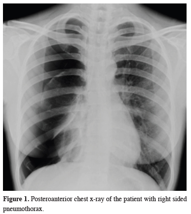
A 45-year-old non-smoker woman was hospitalized for right side pneumothorax (Figure 1) and she was treated with tube thoracostomy.
 Click Here to Zoom |
Figure 1: Posteroanterior chest x-ray of the patient with right sided pneumothorax. |
A thoracic computed tomography (CT) scan taken after the treatment of first attack showed no evidence of intrathoracic abnormalities (Figure 2).
 Click Here to Zoom |
Figure 2: Thoracic CT coronal section image taken after the treatment of first attack. |
One week after the treatment she admitted to our clinic again with recurrent pneumothorax then she underwent right-sided video-assisted thoracoscopic surgery (VATS). Apical wedge resection and partial pleurectomy were performed. During VATS no diaphragmatic pores were detected.
The patient admitted to the hospital with dyspnea and chest pain 1.5 years later. Partial right sided pneumothorax was observed on the posteroanterior chest x-ray. After nasal oxygen therapy the pneumothorax was completely resolved within three days. In the detailed medical history, the relationship between cyclic chest pains and pneumothorax attacks with the menstrual cycle was noticed. Serum CA125 levels were increased moderately (45 U/mL-1). The patient was consulted to the gynecology clinic and GnRH analog treatment was initiated.
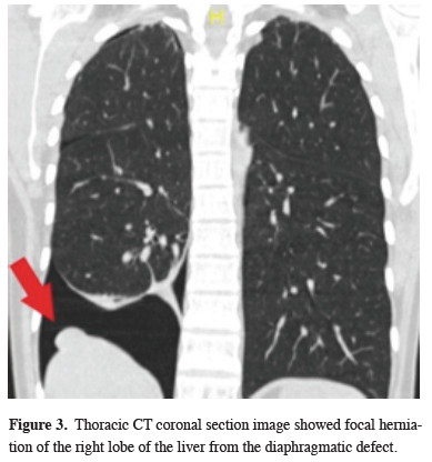
During 1-year follow-up with hormone therapy, there was no chest pain or pneumothorax. Two months after the end of hormonal treatment, the patient complained of chest pain again. The x-ray showed a right pneumothorax thereupon thorax CT was performed. Thorax CT showed a lesion with a size of 22x14 mm adjacent to the right hemidiaphragm suspicious for endometriosis focus or liver parenchyma herniated from diaphragmatic defects (Figure 3).
 Click Here to Zoom |
Figure 3: Thoracic CT coronal section image showed focal herniation of the right lobe of the liver from the diaphragmatic defect. |
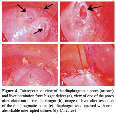
Thoracic magnetic resonance imaging (MRI) was performed for detailed evaluation. Focal herniation of the right lobe of the liver from the diaphragmatic defect was reported in thorax MRI. A new intervention was planned and the patient underwent video-assisted thoracoscopic surgery. Multiple pores were observed in the central tendinous portion of the diaphragm where the right lobe of the liver was herniated from the largest one (Figures 4a-c). The defective area was partially resected and sent for pathologic examination. The diaphragm was repaired with nonabsorbable interrupted sutures (Figure 4d).
 Click Here to Zoom |
Figure 4: Intraoperative view of the diaphragmatic pores (arrows) and liver herniation from bigger defect (a), view of one of the pores after elevation of the diaphragm (b), image of liver after resection of the diaphragmatic pores (c), diaphragm was repaired with non-absorbable interrupted sutures (d). (L: Liver) |
There was no evidence of endometriosis reported on the pathologic examination. The patient was evaluated with gynecology clinic and hormonal therapy (dienogest) was initiated after the surgery. In the postoperative 9 months follow up, the patient had no symptom and no pneumothorax was observed in the chest x-rays.
Written informed consent was obtained from the patient for publication of her data.
Discussion
CP is a recurrent clinical condition that usually occurs one day before or 72 hours after menstruation in premenopausal women. The diagnosis is mostly made clinically and most patients have complaints of typical shortness of breath, and recurrent chest pain at times associated with menstruation [2].CP is mostly associated with thoracic endometriosis. Thoracic endometriosis is defined as the migration of ectopic endometrial tissue into the thoracic cavity, which is the most common extra pelvic location of endometriosis [3]. However positive histopathologic confirmation of thoracic endometriosis is not possible in most of the cases and that is thought to be related to the place where the biopsy was taken randomly [2].
There are a few hypotheses for the pathogenesis of CP. When there is no mucus plug between the atmosphere and the peritoneal cavity during menstruation, the air can pass through the genital system and from the diaphragmatic defects to the thoracic cavity [1]. These diaphragmatic defects may be congenital or acquired and play an important role in pathogenesis. Increased prostaglandin levels during menstruation leading to bronchoconstriction and alveolar rupture or air leakage from endometrial implants on the pleural surface are among other theories [4].
While CP is predominantly on the right side, left-sided and bilateral cases have also been described [1]. Peritoneal fluid containing air and endometrial tissue mainly flows into the right suprahepatic space because of the anatomical difference between the right and left abdominal cavity. This fluid and air pass through the lymphatic pores or existing diaphragmatic defects to the thoracic cavity during respiration. The common occurrence of CP on the right side is explained by this theory [2].
Characteristic lesions of the disease include diaphragmatic perforations and also diaphragmatic or pleural spots or nodules in addition to bullae or blebs. In our case there was no diaphragmatic defect that could be observed in the first operation, however years later more than one defect developed and the liver became herniated. There are only seven cases in the literature with partial liver herniation from diaphragmatic defects and most of them had herniation after recurrence as in our case [5]. The appearance of apical bulla/bleb during the operation may cause the clinicians to approach treatment with the diagnosis of primary spontaneous pneumothorax. A study by Shrestha et al [4] stated that most of the female patients of reproductive age who applied with spontaneous pneumothorax were not questioned about menstrual history at the time of admission to the hospital, all of the patients applied with more than two episodes of pneumothorax, and even two patients had a history of unsuccessful pleurodesis and the diagnosis of catamenial pneumothorax was made mostly retrospectively with intaoperative or histologic findings.
The sensitivity of serum CA 125 level in diagnosis is not clear, Bagan et al [3] emphasized that an increase in serum CA 125 level may be an indicator of endometriosis and may support the indication of videothoracoscopy and hormonal therapy in the early stage.
There is still no clear treatment algorithm for catamenial pneumothorax. Cases with mild symptoms are usually managed with observation, simple thoracocentesis or chest tube, while thoracoscopy, bulla/bleb resection, and pleurodesis are mostly preferred in recurrent pneumothorax [2]. There is no clear consensus regarding the extent of intervention in diaphragmatic lesions [4]. To prevent recurrence and obtain a histological sample, we recommend resection in diaphragm repair and using mesh/graft if necessary. Due to the possibility of recurrence, medical treatment such as GnRH analogs, oral contraceptives and, dienogest are recommended after surgery [6].
In conclusion, with this case, we aim to emphasize that the diagnosis of catamenial pneumothorax should be suspected in patients with recurrent and right-sided pneumothorax, in premenopausal female patients. The entire chest cavity and diaphragm should be fully examined in thoracoscopic examination. Patients with catamenial pneumothorax must be evaluated for endometriosis and a multidisciplinary approach should be provided to the management of the disease, including a thoracic surgeon, gynecologist and, endocrinologist.
Declaration of conflicting interests
The authors declared no conflicts of interest with respect to the authorship and/or publication of this article.
Funding
The authors received no financial support.
Authors’ contributions
GK,BBK; Conceived and designed the analysis, BBK; Collected the data, HK,SGG,BMY; Contributed data or analysis tools, BBK; Performed the analysis GK,BBK; co-wrote the paper.
Reference
1) Bagan P, Le Pimpec Barthes F, Assouad J, Souillamas R, Riquet M. Catamenial pneumothorax: retrospective study of surgical treatment. Ann Thorac Surg 2003; 75: 378-81.
2) Gil Y, Tulandi T. Diagnosis and treatment of catamenial pneumothorax: a systematic review. J Minim Invasive Gynecol 2020; 27: 48-53.
3) Bagan P, Berna P, Assouad J, Hupertan V, Le Pimpec Barthes F, Riquet M. Value of cancer antigen 125 for diagnosis of pleural endometriosis in females with recurrent pneumothorax. Eur Respir J 2008; 31: 140-2.
4) Shrestha B, Shrestha S, Peters P, Ura M, Windsor M, Naidoo R. Catamenial Pneumothorax, a Commonly Misdiagnosed Thoracic Condition: Multicentre Experience and Audit of a Small Case Series With Review of the Literature. Heart Lung Circ 2019; 28: 850-7.


