
Abstract
Materials and Methods: 55 patients were included in the prospective, randomized study with local ethics committee approval. The patients were divided into two groups, and on the postoperative 1st day, pain tape was applied to group I patient and placebo tape similar to pain tape was applied to group II patient. Visual Analogue Scale (VAS) was used for pain assessment, Modified Borg Scale (MBS) for dyspnea assessment, pulmonary function test and six-minute walking test (6 MWT) were used to evaluate pulmonary functions in the preoperative and postoperative periods.
Results: There were 29 patients in group I and 26 patients in group II, with mean age of 59.2 ± 10.4 (18- 75) years. According to the statistical analysis, no difference was found between the groups in terms of age, gender, height, weight, BMI, 6 MWT, MBS, FEV1, FVC, FEV1/FVC values (p > 0.05). There was a difference between the groups in terms of VAS values measured on the 3rd postoperative day (4.65 ± 1.79 vs. 6.57 ± 1.74; p > 0.001). In addition, the postoperative complication rate was different in group I compared to group II (17.2% vs. 30.7%; p = 0.003).
Conclusion: Pain tape application is a reliable and simple method for pain control in the postoperative period in patients undergoing thoracotomy. It can be applied to patients for pain control in the early period, and in this way, the development of postoperative complications can be prevented.
Introduction
Integration of all these analgesia techniques with the pulmonary rehabilitation program can accelerate recovery, prevent postoperative pulmonary complications, and shorten the hospital stay [6,7]. New ones are added to the classical respiratory physiotherapy techniques applied today. One of these new techniques is the application of pain tape, which is known to help in pain management. Pain tape; kinesiology taping technique was developed in the 1970s by Dr. Kenzo Kase to prevent limitations in joint movements and functional activities, which are an undesirable effect in standard tape and tape applications. This tape contains polymer fibers, which has a 60% stretching share, is used to integrate the inflamed areas for any reason, reduce edema, and support circulation. Due to the absence of any pharmacological agents in the tape and its hypoallergenic feature, it provides ease of use in all patients from the pediatric group to the geriatric group [8].
The aim of our study is to investigate the effect of pain tape application on postoperative pain, dyspnea level, and respiratory functions in addition to routine analgesic treatment in patients undergoing thoracotomy.
Materials and Methods
For postoperative analgesia, dexketoprofen (Arveles© 50 mg, UFSA İlaç, İstanbul, Turkey) in 2x1 doses and paracetamol (Parol© 10 mg/mL, Atabay İlaç Fabrika A.Ş., Istanbul, Turkey) 3x500 mg IV was given for all patients. Tramadol hydrochloride IV (Contramal© 100 mg/2 mL, Abdi İbrahim, Istanbul, Turkey) and pethidine hydrochloride IM (Aldolan© 100 mg/2 mL, LIBA laboratories A.Ş., Istanbul, Turkey) were administered when necessary.
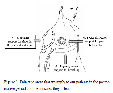
The patients who were taken to the service on the 1st postoperative day were divided into two groups, and pain tape (Kinesio Taping Association International, Albuquerque, NM, USA) was applied to group I (n = 29) patients and a placebo tape similar to pain tape was applied to group II (n = 26) patients. Pain tape taping was applied to the patients on the 1st postoperative day together with the dressing by a physiotherapist who is an expert in the field. The application to be made in the placebo control group was applied by the same specialist physiotherapist to the same areas with the clothing tape in a way that mimics the application of the pain tape. The physiotherapist applying taping did not know whether the tapes were placebos or real tapes. Tape application was made in standardized length, tension, and shape. Three skin areas designated for application, the pain trigger point, were found on the patient by palpation. Y-shaped deltoid/trapezius inhibition on the ipsilateral deltoid/trapezoid and Y- and I-shaped tapings were made on the diaphragm for relaxation and facilitation [9] (Figure 1). The tapes were removed on the day the patient was discharged.
)
 Click Here to Zoom |
Figure 1: Pain tape areas that we apply to our patients in the postoperative period and the muscles they affect. |
In the study, Visual Analogue Scale (VAS) [10] was used for pain assessment, Modified Borg Scale (MBS) [11] for dyspnea assessment, six-minute walk test (6 MWT) [12] and pulmonary function test (PFT) were used to assess pulmonary functions in the preoperative and postoperative periods. VAS and MBS were applied preoperatively and postoperatively on the 1st and 3rd days, 6 MWTs were performed preoperatively and postoperatively on the 3rd day, and PFT was performed at the preoperative and postoperative 3rd month. The specialist physiotherapist and patients who made these evaluations did not know which group they were in.
The inclusion criteria for the study are; patients who have undergone thoracotomy, patients who have not applied a pain tape before the surgical procedure, patients who can cooperate in the postoperative period, patients who give voluntary consent and patients between the ages of 18-80. The exclusion criteria for the study are; patients staying in the intensive care unit for more than 24 hours after surgery, patients with previous thoracic trauma, thoracotomy, or tube thoracostomy, patients who receive psychiatric treatment and cannot cooperate due to the drugs they use, patients who cannot give their own voluntary consent, patients under 18 years of age and patients who develop allergies to the applied pain tape.
Statistical Analysis
Whether the data met the normality assumption was checked using the Shapiro-Wilk test. Mann-Whitney U test was used to test the difference between two groups in terms of ordinal or non-normally distributed continuous variables. Results are expressed as mean value ± SD, or median value and interquartile range (IQR). Bonferroni correction was applied for multiple tests. Number and % values were given for categorical variables. Since the data to be obtained from the study will be subjected to the t-test, considering the power and sample size test based on previous studies in this area, the sample size was calculated as alpha=0.05 and the power of the test was 90%, with a minimum of 25 patients per group. SPSS Windows version 24.0 (Chicago, IL, USA) package program was used for statistical analysis and p < 0.05 was considered statistically significant.
Results
)
Table 1: General characteristics and data of the patients.
There was no significant difference between the groups in terms of FVC, FEV1, FEV1/FVC and PEF values according to the pulmonary function test results performed in the preoperative and postoperative period. Again, although MBS and 6 MWT values performed in the postoperative period were better in group I patients, no statistical difference were found between the groups. When VAS values were evaluated, there was no significant difference between preoperative (1.0 ± 0.88/ 1(2) vs. 0.92 ± 0.97/ 1(2); p = 0.762) and postoperative 1st day values (6.62 ± 1.84/ 7(3) vs. 7.07 ± 1.54/ 7(2); p = 0.323), while VAS values were significantly lower on the 3rd postoperative day in group I patients (4.65 ± 1.79/ 4(3) vs. 6.57 ± 1.74/ 6.5(3); p < 0.001) (Table 2).
)
Table 2: Comparison of the groups in terms of preoperative and postoperative data.
Although hospitalization time (6.5 ± 3.01 days vs. 7.8 ± 3.8 days; p = 0.167) and chest tube removal time (4.9 ± 1.81 days vs. 5.2 ± 2.08 days; p = 0.354) were shorter in Group I, no statistical difference was found. When compared in terms of postoperative complications, it was found to be 17.2% (n = 5) in group I and 30.7% (n = 8) in group II, which was statistically significant (p = 0.003) and the most common complication was atelectasis. (Table 1).
Discussion
In the postoperative period, as after many surgeries, severe pain occurs in approximately 70% of patients after thoracotomy. Pain after thoracotomy is an acute traumatic pain and occurs as a result of stretching of the costovertebral, costotransverse joint ligaments, and posterior spinal muscles [1,2]. After all thoracotomy operations, whether pulmonary resection is performed or not, respiratory muscle strength decreases and respiratory reserve decreases due to the deteriorated respiratory physiology due to the surgical procedure. In addition, postoperative pain restricts diaphragmatic movements by creating phrenic nerve inhibition and causes a decrease in its strength [13]. As a result, breathing becomes shallow, lung volume and capacity decreases, coughing reflex is impaired, causing sputum retention, atelectasis, hypoxemia, hypercapnia and respiratory failure. In addition, age, cardiopulmonary comorbidities, smoking history, variable and low pulmonary function test results, and low exercise capacity increase the risk of postoperative pulmonary complications (PPC) [14-16].
Although developments in postoperative pain management, innovations in anesthesia, and surgery in the last 20 years have reduced general postoperative complications, the most important cause of mortality and morbidity in thoracic surgery is still postoperative pulmonary complications. Factors such as the operation performed, the type of incision and the size of the tissue removed may affect the result, and the rate of PPC can reach 36% [17,18]. In addition, PPC increases the length of stay of patients in the intensive care unit and hospital, and health expenditures [7,19].
To date, various treatment strategies and methods have been developed to reduce the incidence of PPC, such as the detection and prevention of risk factors, improvement of preoperative condition and patient education, intraoperative applications, and postoperative pulmonary care [16-18]. Among these, respiratory physiotherapy has been accepted as a useful practice in preventing pulmonary complications and it has been recommended to be routinely applied by the European Respiratory Society (ERS), European Society of Thoracic Surgeons (ESTS) [20] and American College of Chest physicians (ACCP) [21] due to its functional benefits.
According to the current literature, postoperative pulmonary rehabilitation applied in the early period after thoracic surgery aims; to increase alveolar ventilation, to increase lung volume and capacity, to ensure effective coughing, and to prevent areas under mechanical pressure from going into atelectasis during surgery [13,18]. Although it is stated in the literature that postoperative physiotherapy is necessary and beneficial, there is no standard information about which program should be applied to which patient. The rehabilitation program to be implemented should be planned individually according to the patient’s clinic and needs [7,18]. In our study, we applied the pain tape taping technique, which is a pulmonary rehabilitation application method that has not been investigated and mentioned much in patients who underwent thoracotomy.
The pain tape taping technique is a rehabilitation method that was developed in the 1970s and consists of application. A specially designed elastic tape (Kinesio®) is applied to the patient’s skin to replace the underlying soft tissue areas and achieve therapeutic effects [8]. By causing the skin to rise in the area where the pain patch is applied, it increases the skin and subcutaneous interstitial area, thus increasing circulation and movement in this area. The increase in circulation and movement directs the exudate to the lymphatic pathways, reducing the edema and inflammation and reducing the pain. In addition, the pain tape provides stimulation of mechanoreceptors through the skin, thereby activating the central nervous system [8,22]. In our study, we also used the pain tape taping method, which is easy to apply and does not cause any discomfort to the patient, and we found that the patients in the group in which we applied the pain tape had less pain in the early postoperative period and accordingly, the postoperative complication rate was lower.
The modified Borg scale was developed by the Borg in 1970 to measure the effort expended during physical exercise. It is a scale that is frequently used to evaluate the severity of dyspnea on exertion and the severity of dyspnea at rest [11]. Studies have reported that MBS is a reliable scale for determining the severity of resting and exertional dyspnea and is correlated with respiratory rate and pulmonary function tests. In addition, it is emphasized that MBS is superior to other scales in terms of long-term reproducibility and can be used to predict respiratory reserves of patients. In a study conducted by Akkoca et al, it was found that there was a relationship between the mean dyspnea severity evaluated by patients with MBS and pulmonary function test parameters. In another study conducted by Güneş et al [23,24], it was determined that there was a relationship between the mean dyspnea severity evaluated by patients with MBS and lung functional parameter values. In our study, we used MBS in our patients in the preoperative and postoperative periods to evaluate the perception of dyspnea. Accordingly, we did not observe a statistical difference between the groups, although the MBS values were lower, especially on the postoperative 3rd day, in our patients who were applied pain tape.
Respiratory system functions are always affected during and after surgery. After pulmonary resection, there is an increase in residual volume as a result of both removal of the lung parenchyma tissue and decreased diaphragm and chest wall mobility. While the FEV1 value shows a rapid decrease in the early period; general anesthesia applications cause a decrease in functional residual capacity. This depends on the anaesthetic agents and muscle relaxants used during surgery [20,21]. When the findings related to pulmonary function tests were examined in our study, we found a decrease in the FVC, FEV1, FEV1/FVC, and PEF values measured in both groups of patients in the postoperative period. This decrease was less in group I patients than in group II patients, but we did not observe a statistical difference between the two groups.
The six-minute walk test, developed by Balke in 1963 to measure functional exercise capacity, is a submaximal, indirect cardiovascular physical fitness test. The parameter it tries to measure indirectly is the maximal oxygen consumption, and it measures the distance the person takes in 6 minutes in meters [12]. 6 MWT is easy to perform and evaluate, it does not require equipment. A normal person’s walking distance is 400-700 meters during this time. The six-minute walk test is a widely used approach to evaluate the change in functional exercise capacity of patients in the pulmonary area [13,25]. In a related study in the literature, it was reported that the exercise capacity measured by the 6 MWT improved significantly more than the control group in the 4-week pulmonary rehabilitation program applied to lung cancer patients after resection [13]. In another study conducted by Spruit et al [26] with patients who underwent resection for lung cancer, it was reported that the 6-minute walking distance of the 8-week pulmonary rehabilitation program was significantly better than the control group. Since it was an easily applicable test in our study, we applied 6 MWT to our patients preoperatively and postoperatively. Accordingly, although postoperative 6 MWT values were better in group I patients to whom we applied pain tape, compared to group II patients, we did not detect a statistical difference between the groups.
In conclusion, there is a need for well-programmed and fully implemented pulmonary rehabilitation programs to reduce pain after thoracotomy operations, eliminate pulmonary complications, and return the patient to their preoperative functional life as soon as possible. Considering the clinical benefits, giving importance to simple, cost-effective and low-risk PR practices in thoracic surgery clinics will be important in terms of the success of the surgery, health costs and patient benefits. In this study, we applied pain tape in addition to routine postoperative pain treatment in patients who underwent thoracotomy, and we found that patients felt less pain in the early postoperative period. From this point of view, pain tape seems to be an easy to apply, inexpensive and effective method, and we recommend it to be applied as a part of the pulmonary rehabilitation program in addition to routine pain treatment in patients.
Declaration of conflicting interests
The authors declared no conflicts of interest with respect to the authorship and/or publication of this article.
Funding
The authors received no financial support for the research and/or authorship of this article.
Ethics approval
The study was approved by the Medical Research Ethics Committee of Ondokuz Mayıs University, Faculty of Medicine (KAEK/383).
Authors’ contribution
SGT, BÇ; Conceived and designed the analysis, collected the data, contributed data/analysis tools, performed the analysis, co-wrote the paper, HKÇ; Conceived and designed the analysis, performed the analysis, co-wrote the paper, ZA; Collected the data, co-wrote the paper.