
Abstract
Introduction
Our aim is to share this rare case and remind that PMG should not be forgotten in differential diagnoses in mediastinal masses.
Case Presentation
)
 Click Here to Zoom |
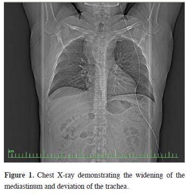
Figure 1: Chest X-ray demonstrating the widening of the mediastinum and deviation of the trachea. |
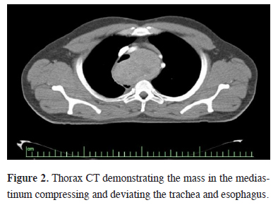
A tracheal shift was evident by palpation during the physical examination. Thorax computerized tomography (CT) revealed a 12x10 cm heterogeneous mass which was located in posterior trachea, adjacent to anterior vertebral column and compressing the esophagus. (Figure 2). Thyroid ultrasonography was normal.
)
 Click Here to Zoom |
Figure 2: Thorax CT demonstrating the mass in the mediastinum compressing and deviating the trachea and esophagus. |
He did not have a thyroid operation previously. Routine laboratory tests including thyroid function tests, beta-human chorionic gonadotropin (B-hcg, 0 mıu/ml), alpha-fetoprotein (αFP, 2.2ng/ mL) and lactate dehydrogenase (LDH, 2,2 U/L) were all in normal ranges. Fiberoptic bronchoscopy revealed a severe external compression trachea.
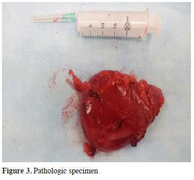
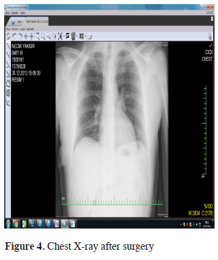
A right lateral thoracotomy is preferred to accomplish a reliable exposure for a posterior mass resection. The mass had no invasions to the adjacent organs and structures and it is resected completely (Figure 3). Histopathologic examination reported macrofolliculer nodular colloidal hyperplasia. Figure 4 shows the postoperative chest x-ray.
)
)
 Click Here to Zoom |
Figure 3: Pathologic specimen |
 Click Here to Zoom |
Figure 4: Chest X-ray after surgery |
Postoperative course was uneventful and he is under follow-up and disease free for 3 years.
Discussion
PMG usually seen in 4th or 5th decades and there is a female preponderance [5]. Although patients are usually asymptomatic and incidentally identified, they may present with symptoms of compression of adjacent organs. Thoracic CT is very sensitive to delineate the nature of the mediastinal masses and their relation to adjacent thoracic structures. Iodine-131 scanning which is used to detect the thyroid tissue in the mediastinal masses, is performed both to investigate the thyroid nature of the mass and to prove that the mass had no relation to cervical thyroid gland.
Thoracic MRI can also be used in addition to thorax CT to plan the surgical approach [6]. βHCG, αFP and LDH levels are essential in the differential diagnosis to distinguish nonseminomatous germ cell tumors from other mediastinal masses. The mass showed a heterogeneous density on CT. Thyroid function tests and βHCG and αFP levels were all normal.
Surgical approach depends on the location of the mass. Median sternotomy, thoracotomy, cervical incision, and thoracoscopy are surgical approaches of PMG resection. In this case we preferred a right lateral thoracotomy due to the localization. The patient was completely symptom free after the resection. After a long term tracheal compression, a postoperative tracheomalacia is a rare but expected complication which was not seen in this case.
In conclusion, this case emphasizes that PMG is a part of differential diagnoses for mediastinal masses despite to its rareness. Thoracotomy is the most common approach to resect mediastinal masses which are greater than 4 centimeter.
Declaration of conflicting interests
The author declared no conflicts of interest with respect to the authorship and/or publication of this article.
Funding
The author received no financial support for the research and/or authorship of this article.