
Abstract
Introduction
Case Presentation
)
)
 Click Here to Zoom |
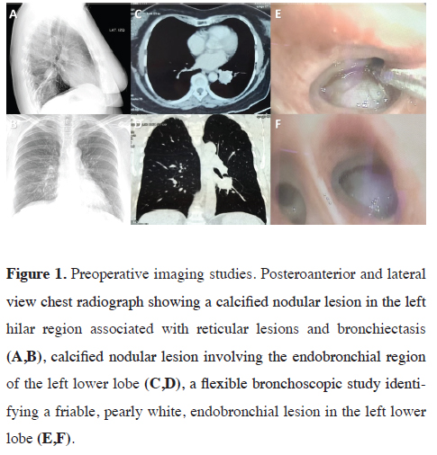
Figure 1: Preoperative imaging studies. Posteroanterior and lateral view chest radiograph showing a calcified nodular lesion in the left hilar region associated with reticular lesions and bronchiectasis (A,B), calcified nodular lesion involving the endobronchial region of the left lower lobe (C,D), a flexible bronchoscopic study identifying a friable, pearly white, endobronchial lesion in the left lower lobe (E,F). |
 Click Here to Zoom |
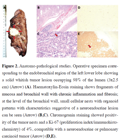
Figure 2: Anatomo-pathological studies. Operative specimen corresponding to the endobronchial region of the left lower lobe showing a solid whitish tumor lesion occupying 98% of the lumen (3x2.5 cm) (Arrow) (A). Haematoxylin-Eosin staining shows fragments of mucosa and bronchial wall with chronic inflammation and fibrosis; at the level of the bronchial wall, small cellular nests with organoid patterns with characteristics suggestive of a neuroendocrine lesion can be seen (Arrow) (B,C). Chromogranin staining showed positivity of the tumor nests and a Ki-67 (proliferation index/immunohistochemistry) of 4%, compatible with a neuroendocrine or pulmonary carcinoid tumor (Arrow) (D,E). |
Discussion
They are usually visualized as solitary pulmonary nodules predominantly in segmental or subsegmental bronchi (80%), in some cases with calcifications, and in up to 75% they are associated with endobronchial lesions as in the present case [4]. Histopathology of TC and AC consist of a uniform organoid growth pattern with cytological features consisting of a moderate amount of eosinophilic cytoplasm with an eosinophilic matrix, with a variety of histological patterns in both, including spindle cell, trabecular, palisade, glandular, follicular, rosette, pink, clear cell, and papillary patterns. Identification and classification can be difficult with immunohistochemistry being important, which points towards a neuroendocrine nature by being positive for synaptophysin, chromogranin, and CD56. However, these can be positive in 30% of cases of lung adenocarcinoma and squamous cell carcinoma, as well as metastatic carcinomas of the breast, prostate, and other sites, and should therefore be considered as differential diagnoses. TTF-1 expression in TC and AC is varied. Most carcinoids stain for cytokeratins, but up to 20% to 25% may be keratin negative. Ki-67 staining shows a low proliferation rate in TC, usually less than 5% while in AC it is higher, usually between 5% and 20%. The proliferation rate may be more useful in small biopsies to separate TC and AC from SCLC, respectively. Ki67 is of limited value in tumors of lung origin, however, it has been estimated that a value > 5% in AC and > 10%, in general, are associated with poor prognosis [6]. The transcription factors TTF-1, CDX-2, and PDX-1 allow differentiation of primary lung origin, being site-specific for lung, gastrointestinal and pancreatic origins, respectively.
Approximately 80 % of low-grade (typical) and 60 % of intermediate-grade (atypical) pulmonary carcinoids express somatostatin receptors by immunohistochemistry and can be imaged using positron emission tomography (PET) or somatostatin receptor scans (OctreoScan) [7]. Typically, 90% are confined to the bronchi and 10% to regional lymph nodes; however, in our case, the nodal staging was negative. As with any neoplastic process, prognosis depends on histological type, stage, and metastatic involvement. Surgical management is of choice and even curative in some cases, there is no standardization, with complete anatomical resection plus mediastinal lymphadenectomy being the most commonly described, however, options range from wedge resection, lobectomy, pneumonectomy or sleeve resection together with mediastinal lymph node study. The latter is controversial, due to the low potential for nodal metastasis of non-active AC and TC described. However, in endocrinologically active tumors a high nodal metastatic potential is presumed, as demonstrated by Seastedt et al who after evaluating 68 cases of pulmonary carcinoids associated with Cushing's syndrome found mediastinal nodal involvement in 37%, with an overall incidence of persistence/recurrence of 16.2% and with a median time to recurrence of 55 months (Range, 18-152 months). He also reported a disease-free time of 12.7 years [8].
Girelli et al analyzed 325 patients with pulmonary carcinoids undergoing complete resection (236 with lymph node involvement and 89 without lymph node involvement), of these 23.6% had TC, 39.3% AC, and 37.1 % large cell neuroendocrine carcinoma. 58.4 % underwent lobectomy, and 21.3 % pneumonectomy, with a mean tumor size of 11-130 mm. Mortality analyzed after 4.0 years of follow-up reached 5.1 %. For TC and AC, the 5-year survival was 89 % and 78 % at 10 years, with a worse prognosis for large cell neuroendocrine carcinoma, 47 % at 5 years, and 41 % at 10 years [9]. Follow-up is carried out taking into account the high potential for recurrence, by clinical and radiological examination. At 3 and 6 months and depending on whether it is TC or AC, annually and semi-annually respectively. Bronchoscopic control may even be indicated in the case of endoscopic resections. These measures should be maintained even for life, with a 10-year survival rate of approximately 90%. The patient underwent follow-ups at 3, 6, and 9 months with no evidence of recurrence [10]. Somatostatin receptor (SRS)-based therapies such as 177-Lu-DOTATATE is an effective and safe option in patients with somatostatin receptor (SRS)-positive (TC)- and (AC)-positive progression. Zidan et al reported in 48 patients after 42 months of follow-up a 33% mortality, a progression-free survival of 23 months, and an overall survival of 59 months [7]. In cases where surgical management is not possible, other therapies such as local radiotherapy should be considered and combined with surgery in cases of AC and Large Cell Neuroendocrine Carcinoma.
In conclusion, it should be remembered that the prognosis and survival of these patients also depend on the histological type of clinical stage, with surgery being the treatment of choice even in locally advanced stages.
Declaration of conflicting interests
The authors declared no conflicts of interest with respect
to the authorship and/or publication of this article.
Funding
The authors received no financial support.
Ethical approval
The present article is approved for the committee ethical
(iO56-94/22) of Department Thoracic Surgery.
Authors' contributions
All authors contributed equally to the idea, data
collection, drafting and final approval of the manuscript.
Reference
This article is an open access article distributed under the terms and conditions of the Creative Commons Attribution (CC BY) license (http://creativecommons.org/licenses/ by/4.0/).