
Abstract
Materials and Methods: Patients who underwent reoperation (with thoracoscopy and / or thoracotomy) and who had a previous operation on the same side were retrospectively evaluated. The indications of the first and second operations, degree of adhesions, need to perform thoracotomy, and keloid and scar formations of the first incision were analyzed.
Results: A total of 22 patients (mean age, 60.1 ± 9.8; range, 19-83 years) who underwent reoperation on the same side were evaluated; 18 of them underwent reoperation due to the recurrence of the first tumor. Hypertrophic scars were observed at the incision site in six (one of them was with keloid formation), advanced pleural adhesions were found in five, and thoracotomy was required in three patients due to severe adhesions. Severe pleural adhesions were observed in 7 of 16 patients without hypertrophic scar.
Conclusions: Postoperative adhesion is a major cause of difficulties and complications during repeat surgeries. More severe pleural adhesions were observed in patients who had hypertrophic scar formation in the previous incision scar, and this observation influenced the clinical approaches.
Introduction
This study aimed to evaluate whether the previous surgical incision scar has predictive value for postoperative adhesion in the thorax.
Materials and Methods
Between January 2017 and October 2022, a total of 22 patients (14 males, 8 females; mean age 60.1 ± 9.8 years; range, 19 to 83 years) who underwent reoperation on the same side were evaluated.
Results
)
Table 1. Patient demographics and clinical characteristics.
We report our experience with two patients with different indications and features.
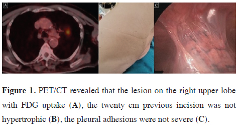
Case 1: A 78-year-old man presented with symptoms of chronic obstructive pulmonary disease (COPD). A 4.6 cm nodule at the left upper lobe was detected with thoracic computed tomography (CT) during the evaluation due to COPD. The 18F-fluorodeoxyglucose (FDG) uptake was 5.3 at positron emission computed tomography (PET-CT) (Figure 1A). The patient had a history of renal cell carcinoma and nephrectomy in 2014. He also had thoracotomy and pleurectomy due to pneumothorax and prolonged air leak on the left side 20 years ago. The incision of the previous thoracotomy was about 20 cm, but the scar was mild without hypertrophy keloid formation on the scar tissue even after the thoracotomy (Figure 1B). Fiberoptic bronchoscopy failed to reveal a final diagnosis. However, we planned to perform a resection (lobectomy) due to suspicion of malignancy. In this patient, we prefer to start the operation with thoracoscopy and then switch to thoracotomy if adhesions associated with the previous operation are severe. Although adhesions are expected to be severe after pleurectomy surgery, adhesions were mild in this patient. The operation could be completed thoracoscopically because of the absence of additional difficulties related to adhesions and previous surgery (Figure 1C). A left upper lobectomy was performed due to tumor localization. Because the lesion was reported as malignant with a frozen section, mediastinal lymph node dissection was added. Postoperatively, no complications occurred, and he was hospitalized for 5 days.
)
 Click Here to Zoom |
Figure 1: PET/CT revealed that the lesion on the right upper lobe with FDG uptake (A), the twenty cm previous incision was not hypertrophic (B), the pleural adhesions were not severe (C). |
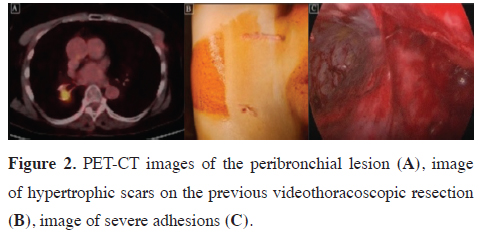
Case 2: A 65-year-old woman was admitted to our clinic from the oncology department because a soft tissue density was detected during follow-up. This right lower lobe peribronchial lesion had a high SUVmax and no suspicion of metastasis in other areas (Figure 2A). The patient had a history of right lower lobe superior segmentectomy for adenocarcinoma by video thoracoscopy 2 years ago. Therefore, the high FDG uptake area was considered as a local recurrence of the first carcinoma. Completion lobectomy was suggested due to no distant metastasis, and her respiratory parameters were good. A severe hypertrophic scar on the previous operation incision was observed during the physical examination (Figure 2B). The operation was started with previous incisions. The Harmonic scalpel (Ethicon Endo-Surgery, Inc., USA) was used for dissection and coagulation of adhesions. Because of severe adhesions, hilar dissection could be started approximately 2h after the initial incision (Figure 2C). The operation was completed with two incisions in a uniportal fashion but took 3h postoperatively, she had an air leak for 2 days and was discharged on postoperative day 4.
)
 Click Here to Zoom |
Figure 2: PET-CT images of the peribronchial lesion (A), image of hypertrophic scars on the previous videothoracoscopic resection (B), image of severe adhesions (C). |
Discussion
Systemic factors (including the female hormone estrogen, hypertension, and hypercytokinemic diseases) were shown to greatly worsen hypertrophic scars and keloid parameters in Ogawa’s meta-analysis [10]. Histologically, localized inflammation has an important role in the formation of hypertrophic skin scars and keloids. Due to inflammation, hypertrophic scars contain inflammatory cells, increased numbers of fibroblasts, newly formed blood vessels, and collagen deposits. Proinflammatory factors, such as interleukin (IL)-1α, IL-1β, IL-6, and tumor necrosis factor-α are upregulated in hypertrophic tissues. Although many agents can be used to prevent postoperative adhesions, most of them are avoided due to side effects. However, corticosteroids and nonsteroidal anti-inflammatory drugs are often used during the postoperative period [11].
Although the relationship between hypertrophic scars and keloids and postoperative abdominal adhesions and their overlapping pathways have not been fully elucidated, Tulandi et al reported that patients with keloids at the wound site had more abdominal adhesions between the uterus and bladder and between the uterus and abdominal wall in repeated cesarean sections [12].
More severe pleural adhesions were observed in patients who had hypertrophic scar formation in the previous incision scar. This observation slightly influenced our clinical approach. Repeat operations were preferred to start with thoracoscopy, and if a problem occurred due to adhesions, then we switched to thoracotomy.
If hypertrophic scar formation is observed at the site of the previous incision in the physical examination of patients, preparations were increased for thoracotomy and the patient was informed about the higher possibility of open surgery. Although surgeries were started with minimal procedures on our patients with or without hypertrophic scar tissue, the conversion rate in open surgery was 2/6 in our patients with hypertrophic scar tissue, whereas this rate was 1/16 in those without hypertrophic scar tissue.
Our study is limited due to the low number of patients and being a retrospective evaluation. Another limitation is that keloids are more prone to occur in some races. However, our study population is not suitable for analyzing the effects of race on adhesions due to geographical reasons.
In conclusion, minimally invasive treatment should be started in patients with previous hypertrophic scar tissue. More caution should be taken in these patients in terms of informing the patients and their relatives preoperatively and preparations for the operating room and surgery. We believe that a prospective multicentric study should strengthen our hypothesis. Another factor that should be considered in a future prospective study is to classify the degree of scar formation and the amount of adhesions based on objective criteria.
Declaration of conflicting interests
The authors declared no conflicts of interest with respect
to the authorship and/or publication of this article.
Funding
The authors received no financial support for the research
and/or authorship of this article.
Ethics approval
This study was approved by the Kartal Lufu Kirdar
Training Hospital Institutional Review Board (May 27,
2024, No: 2024/010.99/4/7).
Authors’ contribution
CT; collected, analyzed and interpreted the patient data,
CT,TC; conceptualized and designed the study, TC;
wrote the paper, collected the literature data, TD; revised
the final version of the manuscript, and co-wrote the paper.
All authors read and approved the final manuscript.
Reference
This article is an open access article distributed under the terms and conditions of the Creative Commons Attribution (CC BY) license (http://creativecommons.org/licenses/ by/4.0/).