
Abstract
Introduction
Case Presentation
)
The tru-cut biopsy result was reported as a lesion containing collagenized stroma and spindle cell areas. Patient’s laboratory findings and tumor markers (CEA = 0.79 ng/mL, CA15-3 = 8.8 U/mL; CA19-9 = 14.1 U/ mL; CA125 = 7.6 U/mL).were within the normal limits.
 Click Here to Zoom |
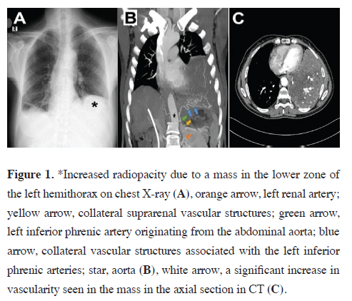
Figure 1: *Increased radiopacity due to a mass in the lower zone of the left hemithorax on chest X-ray (A), orange arrow, left renal artery; yellow arrow, collateral suprarenal vascular structures; green arrow, left inferior phrenic artery originating from the abdominal aorta; blue arrow, collateral vascular structures associated with the left inferior phrenic arteries; star, aorta (B), white arrow, a significant increase in vascularity seen in the mass in the axial section in CT (C). |
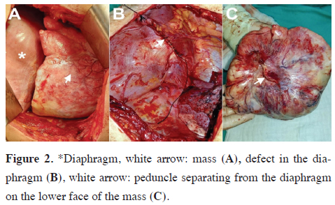
Surgery was performed with posterolateral thoracotomy. A mass was found adherent to the left lower lobe with an approximately 9 cm long peduncle, which was observed intraoperatively to originate from the left hemidiaphragm (Figures 2a,b). The portion of the diaphragm adjacent to the pedicle was resected. The mass was 16×20 cm in size and weighted 1450 gr (Figure 2c). After resection, the left lower lobe was not hypoplastic, filled the lower hemithorax.
)
 Click Here to Zoom |
Figure 2: *Diaphragm, white arrow: mass (A), defect in the diaphragm (B), white arrow: peduncle separating from the diaphragm on the lower face of the mass (C). |
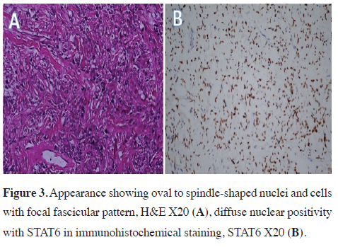
Embolism protocol computed tomography was performed on patient who had complaints of dyspnea on the first postoperative day and pulmonary embolism findings were observed. Low–molecular weight heparin was started. The patient did not show any problems in clinical and radiological follow-ups and hence was discharged. In the postoperative immunohistochemical examination of the patient; neoplastic cells were positive with CD34, bcl2 and diffuse nuclear positive with STAT6 (Figure 3). Pancytokeratin, CD99, DESMIN, CD31, S100 were negative. The findings were reported as compatible with solitary fibrous tumor. Written informed consent was obtained from the patient for the publication of her data.
)
 Click Here to Zoom |
Figure 3: Appearance showing oval to spindle-shaped nuclei and cells with focal fascicular pattern, H&E X20 (A), diffuse nuclear positivity with STAT6 in immunohistochemical staining, STAT6 X20 (B). |
Discussion
In the present case, CECT showed that the mass was supplied by diffuse tortuous collateral arterial structures originating from the abdominal aorta, related to the left phrenic artery and left renal artery. Capsular integrity must be preserved and more careful dissection should be performed in cases with aortic blood supply. Also, lung dissection is recommended after separating the peduncle and controlling the arterial feeding in SFTs, whose blood supply comes from the systemic circulation.
In conclusion, to the best of our knowledge, this is one of the few cases of giant peduncle SFT originating from the diaphragm that was feeding on the abdominal aorta in the English literature. In such cases, en-bloc surgical resection is currently accepted as the universal treatment method. Complete resection provides the best survival in malignant and benign cases. Therefore, surgery is recommended without wasting time after the detection of SFTs originating from the diaphragm.
Declaration of conficting interests
The authors declared no conficts of interest with respect
to the authorship and/or publication of this article.
Funding
The authors received no financial support.
Authors’ contributions
HEC,SEA,VAA,RY,ID: conceived and designed the current
case report, co-wrote the paper, collected the clinical
data. The authors discussed the case under the literature
data together and constituted the final manuscript.
Reference
This article is an open access article distributed under the terms and conditions of the Creative Commons Attribution (CC BY) license (http://creativecommons.org/licenses/ by/4.0/).