
Abstract
Introduction
PPS is usually treated mechanically with tissue expanders or fixed volume prosthesis inserted in the pneumonectomy cavity either by re thoracotomy [2] or minimally invasive surgery [3], repositioning the mediastinum. This approach has proven to be successful in terms of restoring the respiratory and effort capacity of the patients in both subjective (quality of life) and objective (pulmonary function and exercise capacity tests) terms, but may result in significant need for reoperation, morbidity and mortality as well [2]. The overall effect of this kind of surgical correction on gastrointestinal symptoms is unclear.
In this article we present a patient with gastroesophageal reflux disease (GERD) due to post pneumonectomy syndrome, undergoing laparoscopic anti reflux surgery.
Case Presentation
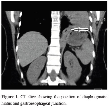
CT scan showed compensatory hyperinflation of the right lung and all mediastinal structures were rotated clockwise to the left hemithorax. Esophagus was stretched and showed crossing over to the left side, exiting the thorax through a larger diaphragmatic hiatus which was caused due to asymmetrical elevation of the diaphragm (Figure 1).
)
 Click Here to Zoom |
Figure 1: CT slice showing the position of diaphragmatic hiatus and gastroesophageal junction. |
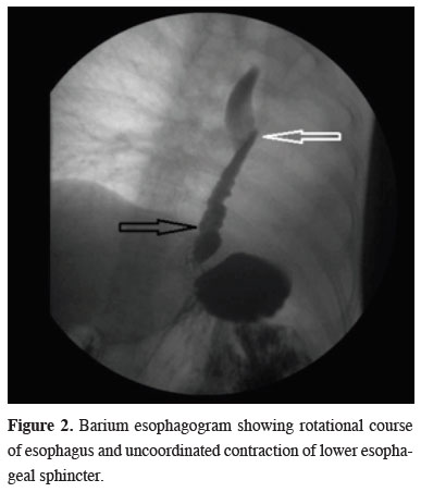
Barium esophagogram showed a normal caliber esophagus with unobstructed passage to stomach, rotational course of the esophagus (Figure 2, white arrow) and uncoordinated contraction of distal esophagus (black arrow) and a small sliding hiatal hernia. pH monitorization and high resolution manometry were concordant with GERD, showing a De Meester score of 70, lower esophageal sphincter (LES) pressure of 28 mmHg and LES length of 2.6 cm. Upper GI endoscopy also confirmed a Hill Class 2 hiatal hernia on retroflexion.
)
 Click Here to Zoom |
Figure 2: Barium esophagogram showing rotational course of esophagus and uncoordinated contraction of lower esophageal sphincter. |
Since the patient had an objective and severe gastroesophageal reflux, we decided to perform a laparoscopic antireflux procedure. Intraoperatively she was found to have a large hiatus, 3 cm hiatal hernia, a smaller and elevated left diaphragm (possibly causing traction to hiatus resulting in enlargement) and severe adhesions between the esophagus and posterior hiatus. Working ports were inserted accordingly, more to the left and superior than normal, adjusting to the patient"s anatomy. Dissection was carried forward towards posterior mediastinum, aorta was seen at the right side of the esophagus. With the help of the posterior mediastinal dissection, an additional 3-5 cm of esophageal length was gained and gastroesophageal junction was restored to its intraabdominal position easily. Diaphragmatic hiatus was repaired posteriorly with two separate 0 silk sutures, a posterior 270-degree fundoplication was also done with two 2-0 silk sutures on both sides. Intraoperatively we didn"t use either mesh or a gastroplasty procedure as repair was possible without any tissue tension.
The patient was able to tolerate clear liquids on postoperative day 1, progressed to soft foods on day 2, and was discharged on day 3 without any complications. She was maintained on soft food diet for 2 weeks, gradually introducing solids thereafter. She remains symptom free in terms of GERD at her 1 year follow up visit and is able to consume a normal diet.
Discussion
Declaration of conflicting interests
The author declared no conflicts of interest with respect to the authorship and/or publication of this article.
Funding
The author received no financial support.