
Abstract
Introduction
Herein we report a rare unusual presentation of EMP in a 67-year-old male with a pleural mass observed on PET, presented with hemoptysis and back pain.
Case Presentation
)
 Click Here to Zoom |
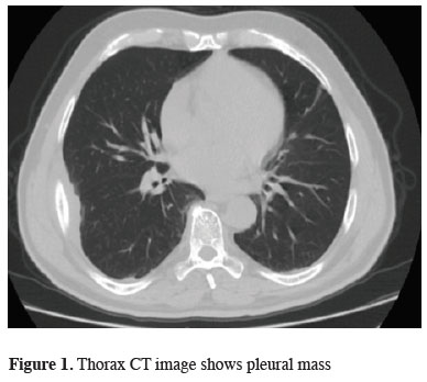
Figure 1: Thorax CT image shows pleural mass |
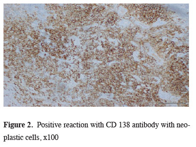
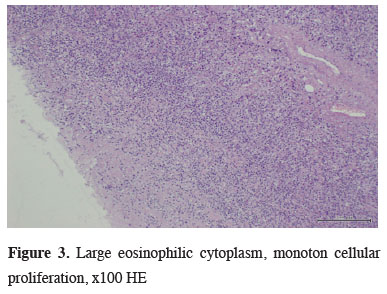
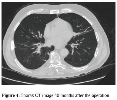
The bronchoscopic examination revealed no endobronchial lesion or abnormal secretions. A pulmonary function test revealed no defect. The whole body positron emission tomography (PET-CT) revealed no other lesion other than right lower pleural mass which had standardized uptake value (SUV) of 5.0. The hemogram, leukocyte differentiation count, and coagulation values were all within normal limits. A CT-guided biopsy, trans-thoracic thin needle aspiration, was performed to conduct a histopathologic diagnosis for the pleural mass, but the result was nonspecific. A biopsy through video-assisted thoracoscopic surgery (VATS) was planned. A 2.5 cm working incision in seventh intercostal midaxillary line and a 11 mm scope port were designed for removing the 3x5 cm pleural mass localized on 5th costal area. The microscopic examination showed a solid mass made up mostly of plasma cells. These tumor cells stained positively for kappa light chains and CD138, but negatively for lambda chains. (Figures 2,3) After confirming that the pleural mass was pleural plasmacytoma, the patient received a series of related evaluations such as serum calcium, urine Bence-Jones protein and plasma electrophoresis for M protein detection. However, all were negative. The skull, spine and pelvis X-ray revealed no osteolytic lesions. The bone marrow biopsy revealed normal patterns of cell distribution. Finally, the patient was referred to the oncology department for radiotherapy. He received external beam radiotherapy 35 Gy in 15-20 fractions over 3-4 weeks which caused radiation pneumonitis. He is on follow-up every 6 months up to date (Figure 4).
)
)
)
 Click Here to Zoom |
Figure 2: Positive reaction with CD 138 antibody with neoplastic cells, x100 |
 Click Here to Zoom |
Figure 3: Large eosinophilic cytoplasm, monoton cellular proliferation, x100 HE |
 Click Here to Zoom |
Figure 4: Thorax CT image 40 months after the operation |
Discussion
Diagnosis of undetermined pulmonary nodular or pleural lesions can be accomplished by transbronchial biopsy, CT guided needle biopsy, as well as surgical biopsy through the VATS or open thoracotomy [6,7]. VATS has been increasingly used for the diagnosis or treatment of undetermined solitary or multiple pulmonary nodules because it is more accurate in diagnosis than needle biopsy and less invasive in treatment than the open procedure [8]. If the result of transthoracic needle biopsy was specific to plasmacytoma, then we would not have performed VATS procedure.
The EMP should be differentiated from other plasma cell tumors. There should be no systemic signs to exclude the possibility of multiple myeloma. In addition, the EMP should also be differentiated from other types of plasma cell tumor, such as reactive plasmacytoma and plasma cell granuloma, or lymphoma such as marginal and immunoblastic subtypes [2,9]. EMP is positive for CD138 and monoclonal cytoplasmic kappa light chain expression of malignant plasma cells [2].
Radiotherapy is the treatment of choice for pleural plasmacytoma because like other plasma cell tumors, EMPs are highly radiosensitive with 80 to 100% of patients successfully achieving local control and with a 50% to 65% ten-year disease-free survival rate [9]. There is no published evidence for the role of adjuvant chemotherapy in the treatment of solitary extramedullary plasmacytoma.
The prognosis of patients with EMP is usually better than those with multiple myeloma. The EMPs, unlike solitary myeloma, advances to multiple myeloma only in a minority of cases. Our patient is observed in each 6 month due to the risk of multiple myeloma formation in patients with plasmacytoma.
We wanted to share this case report to remind our colleagues that plasmacytomas can present as solitary pleural masses and, surgery is used only for the diagnosis. Each PET positive lesion should be explored by minimal invasive methods. As aggressive surgery is not a treatment for pulmonary plasmacytomas, after diagnosis, radiotherapy and oncological follow-up should be appropriate.
Declaration of conflicting interests
The authors declared no conflicts of interest with respect to the authorship and/or publication of this article.
Funding
The authors received no financial support.