
Abstract
Introduction
Patients with lung cancer have been incidentally found to have WT and were misdiagnosed as metastatic disease based on radiologic imaging alone [3]. Here we present an 80-year-old man where PET/CT demonstrated positive cervical lymph nodes for a preliminary diagnosis of a lung tumor. The histopathologic examination of the excised lymph node is reported as a WT.
Case Presentation
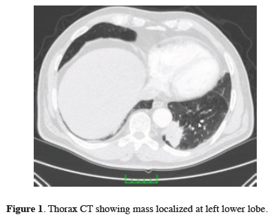
Thorax computerized tomography (CT) showed a 44.5x36.6 mm mass lesion with spicular margins in the posterobasal segment of the left lower lobe close to thoracic aorta at 9th thoracic vertebra level (Figure 1). PET/CT examination demonstrated pathologic 18F- FDG uptake in the defined lesion and bilateral cervical, left hilar and mediastinal lymph nodes. Pathologic uptake was also reported at right iliac bone totally and sacroiliac joint showing sclerotic and lytic changes.
)
 Click Here to Zoom |
Figure 1: Thorax CT showing mass localized at left lower lobe. |
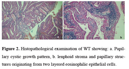
An excisional biopsy of palpable right upper cervical lymph node was performed for tissue diagnosis. Histopathological examination of the lymph node revealed a different tumor growth from lung carcinoma, which contained double row epithelial papillae structures and cystic areas, and central germinal marked lymphoid tissue under the oncocytic epithelium and the pathologic findings were compatible with the WT (Figure 2).
)
 Click Here to Zoom |
Figure 2: Histopathological examination of WT showing: a. Papillary cystic growth pattern, b. lenphoid stroma and papillary structures originating from two layered eosinophilic epithelial cells. |
The patient underwent a transthoracic percutaneous fine needle aspiration biopsy for the left lung mass after a nondiagnostic fiberoptic bronchoscopy, which was reported as non-small cell lung carcinoma (NSCLC) resembling a poorly differentiated adenocarcinoma. Considering PET/CT examination data and comorbidities, histopathologic confirmation of mediastinal lymph nodes were not performed. He was deemed inoperable and directed to the department of medical oncology. The patient died approximately 4 months later after a cerebrovascular accident.
Discussion
The majority of WTs are localized in the parotid gland and in 4-10% of patients, they are multicentric and/or bilateral. It grows very slow and it is painless. The current size doubles approximately in 20 years [9]. Pain and fast growth is very rare and suggests a malign transformation, which was reported as 0.06-3% [10]. 8% of WT is at extraparotid localization, periparotid and upper cervical lymph nodes that are known to be the regions where it is most commonly detected [2]. Rapid growth and/or painful lesions especially in the extraparotid localization, creates serious diagnostic difficulties like in this case.
Routine MR imaging protocol at diagnosis is often insufficient [9]. 18F- FDG PET-CT should be used to show the high glucose metabolism in WT [11]. WTs with serious FED involvement is reported as 6-24% [5]. In this case the SUV max value of lung mass was 10.2, mediastinal lymph nodes were between 3.2 to 4.4 and cervical lymph nodes was between 8.5 to 14, where the later was the value of the excised one (WT). Although this SUV max value was considered to be high for a metastatic lymph node, it is compatible with the serious FED involvement of WT as reported in the literature.
Smoking is accepted as the primary etiologic factor and frequently reported in the literature similar to our patient, who was a heavy smoker. Kotwall et al reported that WT is seen eight times more in smokers compared with non-smokers [12]. As smokers have an increased risk for lung cancer, it is not surprising that those with WT are also have a risk of lung cancer.
In the English literature WT and lung cancer coexistence is very rarely reported. Dua et al [13] reported a case with bilateral synchronous and multifocal WT mimicking metastasis from lung cancer, Arora et al [14] reported 3 cases, and Thomas et al [15] reported 2 cases. Although there has been no proven association, a retrospective study by White et al [3] reported that, among 144 patients with WT, 24 (19%) had a concomitant lung cancer, the most common being NSCLC. They suggested that given the association between WT and lung cancer, earlier recognition of these benign lesions might potentially facilitate earlier diagnosis of lung malignancies [3].
As a conclusion, Warthin's tumors that are found at an extraparotid localization might cause a diagnostic dilemma, when they show intense FGD uptake. An appropriate tissue diagnosis should be performed for the misinterpretation of a metastatic disease. Foremost, it would be essential to identify patients with WT, as an association with lung cancer was suggested.
Declaration of conflicting interests
The authors declared no conflicts of interest with respect to the authorship and/or publication of this article.
Funding
The authors received no financial support.