




2Department of Thoracic Surgery, Diskapi Yildirim Beyazit Education and Resarch Hospital, Ankara, Turkey DOI : 10.26663/cts.2018.00012
Summary
Pulmonary alveolar proteinosis (PAP) is a rare disease characterized by accumulation of material in the alveoli of the lungs. It is generally diagnosed in middle-aged men. The most common complaints are cough and dyspnea. Characteristic radiological findings of PAP include nodular infiltration with a tendency to merge and ground glass density, septal thickening and resemblance to paving stone. Typical findings include periodic acid-Schiff (PAS) stain positive materials in the alveolar proteinosis, milky appearance of bronchoalveolar lavage (BAL), interstitial opacities, thickened interlobular septa, and patchy central or peripheral ground-glass opacities in the lung parenchyma in the chest CT. The most effective proved treatment is whole lung lavage. The aim of the treatment in PAP cases is to restore the alveolar gas exchange by removing excess surfactant accumulated in the alveoli.Introduction
Pulmonary alveolar proteinosis (PAP) is a rare disease characterized by accumulation of material in the alveoli of the lungs, which could take primary and secondary forms [1]. It is generally diagnosed in middle-aged men [2]. Typical findings include periodic acid-Schiff (PAS) stain positive materials in the alveolar proteinosis, milky appearance of bronchoalveolar lavage (BAL), interstitial opacities, thickened interlobular septa, and patchy central or peripheral ground-glass opacities in the lung parenchyma in the chest CT. The most effective proved treatment is whole lung lavage [3].Here we present a case of pulmonary alveolar proteinosis, admitted with symptoms of cough and progressive dyspnea, diagnosed with BAL and had atypical scan results, along with a review of literature.
Case Presentation
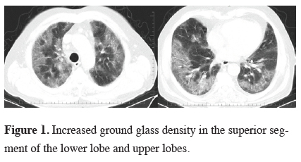
A 70-year-old man admitted to our clinic with complaints of cough and progressive dyspnea of five months duration. According to his medical records, he was not able to meet his basic daily needs and walk without support due to dyspnea. In addition to non-productive cough and dyspnea, he suffered from loss of appetite and fatigue. The findings on physical examination included cyanosis in lips, intercostal recession, decreased breath sounds and low levels of rhonchus. Oxygen saturation was measured as 75% using non-invasive methods.He was hospitalized and empiric antibiotherapy, inhaler and parenteral corticosteroids, diuretic treatment was started. Chest CT showed increased ground glass densities at periphery of both lung parenchyma, particularly in the superior segment of the lower lobe and upper lobes (Figure 1).
)
 Click Here to Zoom |
Figure 1: Increased ground glass density in the superior segment of the lower lobe and upper lobes. |
Despite nasal oxygen support, oxygen saturation level was 82%. Since the case did not have fever and WBC count was as 8300/mm3, no infection was suspected. The laboratory findings revealed hemoglobin level as 13.4g/dL, glycosylated hemoglobin as 8.5, CRP > 32mg/L, albumin as 2.7g/dL and protein in urine as 3+. Since there was no finding of hemoptysis or anemia, alveolar hemorrhage diagnosis was not suspected. No coronary failure was detected in the cardiology scan performed in the cardiology clinic. Fiberoptic bronchoscopy did not reveal any finding of infection, inflammation or malignancy. The right lung was lavaged with 30mL of isotonic saline solution, which showed intensive infiltrative lesions in chest CT. The lavage specimen was in milky appearance. He reported that his complaints was relieved following bronchoscopy. However, due to a decrease in the oxygen saturation to 65% in the following hours, CPAP support using a mask is administered.
On the 2nd day of his hospitalization, pathological examination of the lavage specimen revealed that it is PAS stain positive. He was diagnosed as pulmonary alveolar proteinosis (PAP) and a whole lung lavage was planned. An informed consent was obtained. A positive end expiratory pressure was performed for the following three days, however, the oxygen saturation did not increase and he was supported by mechanical ventilation on CPAP mode. Same day he was intubated and underwent an urgent lung lavage. Following a double lumen selective intubation, 200mL serum physiological with approximately 36 °C was poured into the right lung. During the aspiration of the fluid, a sudden cardiac arrest occurred and resulted in mortality despite resuscitative maneuvers.
Discussion
Pulmonary alveolar proteinosis is a rare disease characterized by progressive accumulation of surfactant, phospholipid and proteins in alveoli and terminal airways leading to impairment of gas exchange. It occurs in three forms including idiopathic (primary), secondary and congenital [4]. Among the causes of secondary form are exposure to heavy inorganic dusts such as silica, titanium, aluminum, hematological and oncological diseases such as leukemia and lymphoma and etiological factors such as HIV and Pneumocystis carinii [5].PAP usually affects adults aged 20-50 years. It is a rare disease which could be asymptomatic and which could result in mortality. The most common complaints are cough and dyspnea. Other symptoms include fatigue, weight loss, fever, pleuritic chest pain and hemoptysis. Initial complaints of our case were progressive dyspnea and cough.
Characteristic radiological findings of PAP include nodular infiltration with a tendency to merge and ground glass density, septal thickening and resemblance to paving stone [6]. Similar findings can be found in hypersensitivity pneumonia, lung edema, alveolar hemorrhage, radiation pneumonia and sarcoidosis [7]. According to the literature, PAP predominantly affects upper and lower parts of lungs and peripheral lung remain protected [8]. However, in our case lesions were also intensive in the periphery.
In cases of PAP, respiratory function tests reveal restrictive respiratory function disorder and mid-level decrease in CO diffusion test [9]. In our case, respiratory function test could not be performed because of advanced dyspnea. However, arterial blood gas analysis showed hypoxemia and increase in shunt fraction.
In cases of PAP, BAL fluid typically has a white-opaque appearance. Analysis of the fluid reveals inflammatory cell, alveolar macrophage, extracellular eosinophils materials and PAS positive material abundant in lipid and protein [6,8,10]. The pathological findings in our differential diagnosis were ruled out with anamnesis and examination. The diagnosis was made based on the typical appearance of lavage fluid and cytological examination of the fluid as PAS positive.
PAP treatment is often decided according to the progression of the disease, presence of infections and general clinical condition of the patient. 20% of the PAP cases can recover spontaneously and mortality in primary form is below 10% [9,11]. In cases with solitary lung opacities, resolution could develop from 3 to 9 months [12]. The aim of the treatment in PAP cases is to restore the alveolar gas exchange by removing excess surfactant accumulated in the alveoli. Despite studies on other treatment methods including systemic steroids, proteinosis, mucolytics, the standard accepted treatment method is still whole lung lavage. In a study conducted with PAP cases, symptoms were alleviated and radiological findings improved in the 52% of the 51 cases that underwent lavage and 94% of the 17 cases that were followed up [13]. There are reports arguing that approximately one third of the cases spontaneously recovered. However, it is also reported that pulmonary fibrosis developed in approximately one third of the cases despite repeating whole lung lavage [6]. The main treatment criteria include PaO2 < 65 mmHg, P (A-a) O2 > 40 mmHg, shunt fraction > 12%, and dyspnea with minimal effort [9].
Our case’s treatment failure depends on its diagnoses was done at the end stage. Desaturation, cyanosis and intercostal retractions at arrival were indicators of bad diagnosis. Identification processes such as laboratory investigations and invasive procedures fulfilled at first day, pursuit of pathology obtained on the second day, and it was planned to wash lung on the third day. The day when the lung lavage was planned, the patient was desaturated and ventilated on CPAP mode by mechanical ventilator for three hours and because hypoxemia persisted the patient had intubated electively. After intubation, lung lavage had completed. Intubation had performed selectively with double lumen tube accompanied by bronchoscopy. In process, 200mL 0.9% NaCl was given and aspirated, so no fluid remained in the lung. We do not think that arrest depend on neither fluid nor process time.
Lung lavage for the treatment of PAP was first proposed by Ramirez-Rivera in the 1960’s [14]. Nowadays lavage treatment is conducted under general anesthesia with approximately 60L serum physiological and using lung ventilation via double lumen endotracheal tube [12].
It announced that PAP patients who have bad general status, need of ventilation, have good results with extracorporeal membrane oxygenation (ECMO) support lung lavage [15]. However, our center has not ECMO background so we could not support the patient with ECMO. Because of the patient’s instability, the patient could not transferred any other center. The deteriorated arrival of the patient to the hospital, fast progressive behavior of the disease, such cases has a high mortality risk. Although the complaints of our case started five months before and progressively increased, no diagnosis could be done. This might have been because of the rarity of PAP cases and radiological findings showing similarities with other lung diseases.
As a conclusion, two characteristics of our case make it different from other reported cases. First of all, it did not present in age 20-50 years and secondly lung periphery has been affected as much as other parts of the lung. As a take home message we want to emphasize that PAP, which is seen in young adult age group, could be seen in older ages and radiological patterns could show differences.
Declaration of conflicting interests
The authors declared no conflicts of interest with respect to the authorship and/or publication of this article.
Funding
The authors received no financial support.
Reference
1) Prakash UB, Barham SS, Carpenter HA, Diners DE, Marsh HM. Pulmonary alveolar phospolipoproteinosis: Experiences with 34 cases and a review. Mayo Clin Proc 1987; 62: 499-518.
2) Wang BM, Stern EJ, Schmidt RA, Pierson DJ. Diagnosing pulmonary alveolar proteinosis. A review and update. Chest 1997; 111: 460-6.
3) Wasserman K, Mason GR. Pulmonary alveolar proteinosis. In: Murray JF, Nadel JA (eds). Textbook of Respiratory Medicine. 2nd ed. USA: Saunders, 1994: 1933-46.
4) Persson A. Pulmonary alveolar proteinosis. In: Fishman AP (ed). Pulmonary diseases and disorders. New York: Mc Graw Hill Book Company, 1998: 1225-30.
5) Selimoğlu Şen H, Dallı A, Önder Ö. F, Abakay A, Fırat U, Eren Ş, et al. Pulmonary alveolar proteinosis. Dicle Med J 2011; 38: 234-8.
6) Menard KJ. Whole lung lavage in the treatment of pulmonary alveolar proteinosis. J Perianesth Nurs 2005; 20: 114-26.
7) Blumenthal NP, Miller WT, Kotloff RM. Radiographic pulmonary infiltrates. AACN Clin Issues 1997; 8: 411-24.
8) Maygarden SJ, Iacocca MV, Funkhouser WK, Novotny DB. Pulmonary alveolar proteinosis: a spectrum of cytologic, histochemical, and ultrastructural findings in bronchoalveolar lavage fluid. Diagn Cytopathol 2001; 24: 389-95.
9) Ulukavak Çiftçi T, Şipit T, Apaydın Z, Çoban G, Oğuz D. Pulmonary Alveolar Proteinosis (PAP) (Due to a Case). Solunum Hastalıkları 2001; 12: 71-4.
10) Goldstein LS, Kavuru MS, Curtis-McCarthy P, Christie HA, Farver C, Stoller JK. Pulmonary alveolar proteinosis: clinical features and outcomes. Chest 1998; 114: 1357-62.
11) Kaçar N, Ünsal Ş, Çakan A, Tuksavul F, Ermete S, Güçlü SZ. Pulmonary Alveolar Proteinosis (case report). Tüberküloz ve Toraks Dergisi 2000; 48: 162-6.
12) Taş D, Demirer E, Kunter E, Küçükodacı Z, Ayten Ö, Okutan O, et al. Pulmonary Alveolar Proteinosis: Report of two cases. Solunum 2011; 13: 182-6.
13) Asamato H, Kitaichi M, Nishimura K, Itoh H, Izumi T. Primary pulmonary alveolar proteinosis. Clinical observation of 68 patients in Japan. Nippon Kyobu Shikkan Gakkai Zasshi 1995; 33: 835-45.