
Abstract
Introduction
Case Presentation
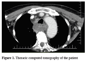
The case was consulted with otorhinolaryngology (ORL) clinics with the pre-diagnosis of deep cervical infection. His cervical thoracic computerized tomography (CT) revealed an abscess formation in the retrotracheal space and he was hospitalized to the ORL clinics with the diagnosis of deep cervical infection. Emergency abscess drainage was performed through oral cavity under general anesthesia at the same day by ORL clinics. Methicillin resistant Staphylococcus epidermidis (MRSE) was found in the culture results of the abscess material and the case was consulted with the infectious diseases clinics. An antibiotherapy consisting Vancomycine 4x500 mg IV, Imipenem 4x500 mg IV and Metronidazole 2x500 mg IV was administered. He was consulted with our clinics at the 4th post-operative day because of ongoing fever and leukocytosis. His physical examination revealed tenderness in the cervical region on palpation. His lung auscultation was normal. Thorax CT revealed mediastinal air fluid levels compatible with DNM, starting from cervical region down to the sub-carinal space especially on the right side of mediastinum (Figure 1).
)
 Click Here to Zoom |
Figure 1: Thoracic computed tomography of the patient |
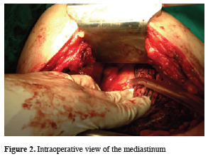
Patient was transported to the operating theater immediately in order to perform an urgent mediastinal debridement with the pre diagnosis of DNM. General anesthesia was performed using a double lumen intubation tube. Videothoracoscopic exploration was performed through the right 5th intercostal space (ICS) via one 10.5 mm thoracoport. Pleural effusion was found and samples were obtained. However, patient’s age and his irregularly treated COPD with infective status caused his blood oxygen saturation levels drop during surgery and patient did not tolerate single lung intubation. Thus, videothoracoscopy was converted to posterolateral thoracotomy through the 5th ICS. Serratus anterior muscle was spared during the procedure. Intraoperative exploration revealed an abscess formation at 4-5 cm superior to the azygo-caval junction protruding into the thoracic cavity just anterior to the esophagus (Figure 2).
)
 Click Here to Zoom |
Figure 2: Intraoperative view of the mediastinum |
The yellowish white colored dense fluid content of the abscess was drained and samples were collected. The abscess cavity was further explored. The cavity reached up to 3-4 cm superiorly into the paravertebral space and 3-4 cm inferiorly until carina. These additional sites were thoroughly debrided as well. Patient was extubated in early postoperative period and was transferred to the intensive care unit (ICU). Mediastinal pus collection was evaluated with daily intravenous contrast enhanced thorax CT. The leukocyte count on the first postoperative day was 14,900/mm3. Patient was transferred to our clinic at the postoperative second day as his general condition improved, and there were no additional mediastinal collection or air-fluid levels on thorax CT. Chest tubes were removed (apical on the 5th, basal on the 7th postoperative day) as the drainage from the chest tubes decreased and the color of the drained fluid became serous. The fever also resolved. The microbiological culture results of the pleural effusion and abscess material obtained during surgery revealed no bacteria attributable to the broad-spectrum empirical antibiotherapy he was receiving. He was discharged on the 10th postoperative day as his physical examination revealed bilateral normal lung auscultation findings with normal posterior-anterior (PA) chest x-ray and blood tests revealed diminished leukocytosis. His outpatient clinics examination one week after he was discharged was normal. The thorax CT on postoperative third month revealed no abnormal findings in the mediastinum.
Discussion
DNM is treatable with a broad-spectrum antibacterial treatment and early surgical drainage. The etiological microorganisms that are isolated in DNM patients usually consist of Streptococcus, Bacteroides, Staphylococcus, Peptostreptococcus, Fusobacterium, Propionibacterium, Stenotrophomonas, Pseudomonas species that are aerobic and anaerobic bacteria and fungi of Candida species [5]. Multiple microorganisms were reported to be isolated in 29% of the DNM patients [6]. We isolated MRSE in our case. The culture and antimicrobial susceptibility tests for the MRSE revealed that the isolated bacteria was susceptible to Vancomycine but in order to achieve a broader spectrum for any additional microorganisms that could not be identified we continued administering Imipenem and Metronidazole to the patient.
It was also reported that especially immunodeficiency secondary to diabetes mellitus (DM) is an important risk factor in the development of DNM [7]. We believe that the absence of DM in our case contributed to the good prognosis.
In addition to these, the most important prognostic factors in DNM were advocated as early diagnosis and prompt treatment [8]. The most important factor in achieving early diagnosis in DNM is clinical suspicion. Clinical suspicion attributable to the long lasting sore throat and fever in our case helped us establishing the deep cervical infection. Early diagnosis and prompt surgical drainage of the cervical and mediastinal regions and early administration of broad-spectrum antibacterial treatment affected the patient’s clinical status in a good way.
As conclusion, the most important factor affecting the morbidity and mortality in DNM is early diagnosis and prompt treatment with early aggressive surgical drainage. Broad-spectrum antimicrobial treatment tailored according to the culture results of abscess material will significantly improve the prognosis in DNM patients.
Declaration of conflicting interests
The authors declared no conflicts of interest with respect to the authorship and/or publication of this article.
Funding
The authors received no financial support.