
Abstract
Introduction
Case Presentation
)
 Click Here to Zoom |
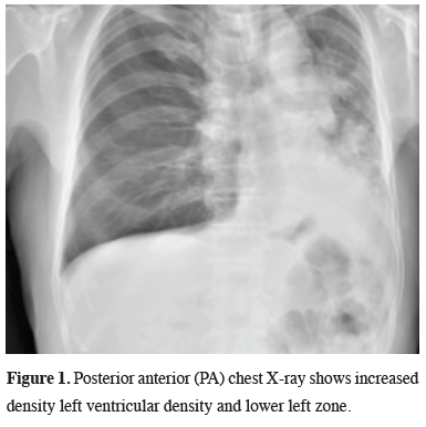
Figure 1: Posterior anterior (PA) chest X-ray shows increased density left ventricular density and lower left zone. |
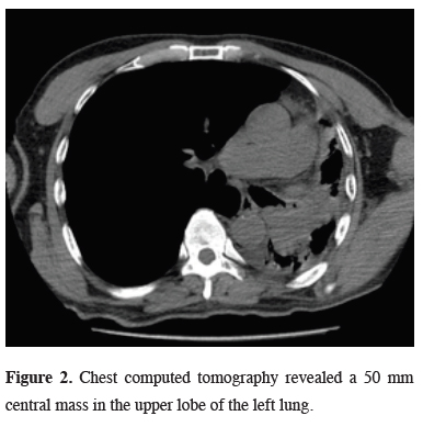
Chest computed tomography (CT) and positron emission tomography (PET/CT) reported a 50 mm central mass (SUVmax: 13) in the left upper lobe of the lung (Figure 2).
)
 Click Here to Zoom |
Figure 2: Chest computed tomography revealed a 50 mm central mass in the upper lobe of the left lung. |
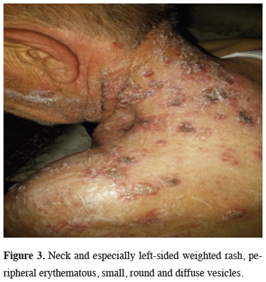
Fiberoptic bronchoscopy (FOB) examination revealed an endobronchial lesion in the left upper lobe bronchus and biopsy was reported as squamous cell carcinoma. Lung function tests were within normal limits (FEV1: 2.1 L). The patient was found to have suspicious mucocutaneous lesions (Figure 3) and no pathology was detected in the biopsy of these lesions. Herpes zoster was diagnosed clinically.
)
 Click Here to Zoom |
Figure 3: Neck and especially left-sided weighted rash, peripheral erythematous, small, round and diffuse vesicles. |
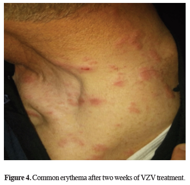
Analgesic and acyclovir treatment lasted for fourteen days. Common erythema after two weeks of VZV treatment is seen in Figure 4.
)
 Click Here to Zoom |
Figure 4: Common erythema after two weeks of VZV treatment. |
Mediastinoscopy was performed and lymph nodes (2R-4R-7) were primarily sampled. Left pneumonectomy was performed with thoracotomy on preoperative lymph node reactive residence. The case was pathologically staged as T3N0M0 Stage IIB. The patient was discharged uneventfully with no recurrences at 18 month follow-up.
Discussion
Herpes zoster can usually be diagnosed clinically. However, the diagnosis can be confirmed by the vesicle exacerbations seen with the viral cultures of the vesicular juice, which reveal the multicellular giant cells. Histology reveals inflammation and neuronal loss in the corresponding ganglia in affected dermatomes [4]. In our case, lung cancer and VZV were detected in the same hemithorax region. It shows that VZV may be a secret cancer marker. Evidence for subtype cancers suggests a strong association between herpes zoster and occult hematological cancer. In an anthropological review published in 1995, the relationship between herpes zoster and the diagnosis of subsequent malignancies was examined. Based on only two small basic publications of this review, the authors of the review concluded that there was no increased risk of cancer within herpes zoster patients [5]. Various biological mechanisms explaining the relationship between cancer and herpes zoster has been previously suggested [6]. Findings of VZV in our patient may be related to lung cancer. First, carcinogenesis can induce immunodeficiency, which causes the herpes zoster to burst in the preclinical asymptomatic stage. In particular, it may lead to impaired number and function of B-cells and T-cells positively associated with hematological cancers and these cancers. The risk of hematological cancer increases even 5 to 10 years after the herpes zoster and this immunosuppressive effect indicates that diagnosis of cancer can be detected several years ago [7]. Cellular immune function is firstly important for suppressing varicella-zoster virus replication. Secondly, cellular immune function is crucial to suppress varicella-zoster virus replication and carcinogenesis. For this reason, herpes zoster may be a marker of impaired immune surveillance in the host and in this case causes cancer development. For example, immunosuppression in autoimmune diseases can increase the risk of herpes zoster and some cancers [8]. Thirdly, it is possible that recurrent antigenic stimulation resulting from subclinical and eventual activation of the clinical varicella zoster virus leads to exacerbation of precancerous genetic lesions or triggering neoplastic transformation. Various herpesviruses act directly on tumor-induced disease through inflammation, decreased immunity and immunosuppression. A direct carcinogenic effect of herpes zoster may also explain the cancer development in previous areas affected by herpes zoster [9].
Shingles can occur with local trauma or procedures. The majority of the reported cases develop in the same dermatome as the previous trauma. However, several cases have been reported about where the rashes are located in the real incision area. These cases show that shingles events developed around an incision site may mimic other rash causes. There were very few reports on the surgical incisions of the patients that developed shingles. While Godfrey et al reported that the thoracic surgeon for scoliosis was a zona-developing disease at the surgical incision site, Choi et al published a case report of a patient who developed a herpes zoster within the last face surgery scar [10]. Thomas et al. published a case-control study showing that the risk of shingles increased for one month following trauma, 39 studies in this study identified an increased risk for breast cancer during the next two years of radiotherapy [11]. However, Gadient et al. studied the time interval between the process and the development of the rash [12]. The purpose of treating shingles is to reduce the duration of symptoms of the disease, reduce the risk of transmission and prevent post-herpetic neuralgia. Post-herpetic neuralgia is a syndrome that develops in about 10% to 15% of cutaneous hypersensitivity and neuropathic pain and is particularly difficult to treat [13]. Our case was operated after VZV treatment.
Only a few studies have examined the interactions with age and time after the diagnosis of malignancy [14]. As a conclusion, the relationship between herpes zoster and malignancies should be kept in mind.
Declaration of conflicting interests
The authors declared no conflicts of interest with respect to the authorship and/or publication of this article.
Funding
The authors received no financial support.