
Abstract
Materials and Methods: We designed a retrospective study examining the utility of NLR and PLR in estimating trauma severity in sternum fracture patients who admitted to our hospital between January 2013 and April 2018. Patients were divided into two groups: isolated sternum fracture group (36 patients with sternal fracture and without any complications in thorax or other system and/or only 1 or 2 rib fractures) and complicated sternum fracture group (19 patients with sternal fracture and with associated complications in thorax or other systems). Injuries were not graded but referred as severe according to the associated complications in thorax or other systems. The key clinical parameters, including NLR and PLR were compared among the groups.
Results: Based on the ROC curve, the best NLR cut-off value to predict complicated sternum fracture group patients was 2.1, with 88.9% sensitivity and 28% specificity, whereas the best PLR cut-off value was 123.8, with 72% sensitivity and 68% specificity.
Conclusions: The results may indicate a clinically useful marker that can be easily and reliably measured from a blood sample to predict outcome of trauma patients with sternal fractures.
Introduction
Stimulation of a variety of inflammatory mediators takes place immediately after trauma. Although the level of CRP has been used in clinical practice for many years, it is relatively non-specific and many clinical studies have shown that there is no correlation between the serum CRP and the severity of injury or prediction of survival in poly traumatized patients. In this manner several studies have been published addressing the search for a clinically useful marker that can be easily and reliably measured from a blood sample [4].
The neutrophil-to-lymphocyte ratio (NLR) and platelet-to-lymphocyte ratio (PLR) have been defined as novel markers of inflammation and thrombotic events which can be easily measured from the complete blood cell count (CBC). NLR possesses diagnostic value in certain pathologies characterized by systemic or local inflammatory response such as acute coronary syndrome, non-ST myocardial infarcts, ischemic and hemorrhagic strokes, pulmonary embolisms, and several types of cancer [5-7]. PLR is currently considered to be a more sensitive marker and also assumed as a prognostic factor in breast cancer, and ovarian and colorectal cancers [8].
Although the NLR and PLR are cheap and widely available biomarkers, literature regarding their usage in trauma patients is scarce. It seemed to us interesting to evaluate the prognostic value of the NLR and PLR for trauma patients. In this manner, we selected a small group of trauma patients with sternal fractures and aimed to investigate their correlation with the serum NLR and PLR. To the best of our knowledge, this is the first study investigating the clinical value of these cheap and widely available biomarkers in trauma patients.
Materials and Methods
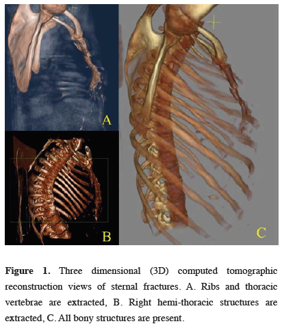
Injuries were not graded but referred as severe according to the associated complications in thorax or other systems. An ISF was considered as a sternal fracture without other thoracic or concomitant injuries except two or less rib fractures. A complicated sternal fracture (CSF) was regarded as a severe injury including a sternal fracture with associated other thoracic (more than two rib fractures) or concomitant injuries. Patients were divided into two groups: isolated sternum fracture (ISF) group (36 patients with sternal fracture and without any complications in thorax or other system and/or only 1 or 2 rib fractures) and complicated sternum fracture (CSF) group (19 patients with sternal fracture and with associated complications in thorax or other systems) (Figure 1).
)
 Click Here to Zoom |
Figure 1: Three dimensional (3D) computed tomographic reconstruction views of sternal fractures. A. Ribs and thoracic vertebrae are extracted, B. Right hemi-thoracic structures are extracted, C. All bony structures are present. |
Patient demographics (age and sex), cause of trauma, type of care (inpatient, outpatient), laboratory results (including NLR and PLR from complete blood count), length of stay data were gathered and analyzed. Imaging investigations included x-ray (lateral chest x-ray or sternal view x-ray) and chest CT in all cases. The SF was diagnosed by x-ray or chest CT. An electrocardiogram (ECG) was also performed. An Echocardiogram (ECHO) was performed only in patients with pathological ECG or suspected pericardial effusion on chest CT. Investigations including cardiac enzymes such as creatine phosphokinase (CPK), myocardial branch of creatine phosphokinase (CKMB) and high sensitive troponin (Hs-Tn) were also recorded.
Pericardial effusion, myocardial contusion and vascular pa¬thologies were considered as abnormal ECHO findings. ST segment changes were accepted as abnormal ECG findings. Hs-Tn and CK-MB tests were evaluated; levels above 14 ng/L of Hs-Tn and/or above 5% of CK-MB (percentage of total CK) were con¬sidered as cardiac enzyme elevation.
Laboratory parameters
The laboratory results were evaluated according to the first venous blood samples taken on admission to the emergency department. Samples were analyzed within 1 h of collection using an automated blood cell counter (Coulter® LH 780 Hematologic Analyzer, Beckman Coulter Inc. Brea, USA). Reference values were 2.1–6.1×10³/mm3 for neutrophils, 1.3–3.5×10³/mm3 for lymphocytes, 156– 373×103/mm3 for platelets. The NLR was calculated as the ratio of the neutrophil count to the lymphocyte count, and the PLR was calculated as the ratio of platelet count to lymphocyte count.
Statistical Analysis
Statistical analysis was performed using IBM SPSS Statistics version 16.0 software (SPSS Inc., Chicago, IL). Median and inter quartile range (IQR) were used as descriptive statistics for continuous variables. To compare qualitative variables, Fisher’s Exact Test was used.
Mann–Whitney U test was used for analysis of non-parametric continuous data. p value < 0.05 was considered as statistically significant.
Results
)
)
Table 1: Baseline characteristics of the study population.
The most common associated pathology was rib fracture (36.3%) in 20 patients with SF. Other intrathoracic injuries associated were; pneumothorax in one (5.2%) patient, hemopneumothorax in one (5.2%) patient, pulmonary contusion in two patients (10.5%) and hemothorax in two patients (10.5%) in CSF group. The most common extrathoracic associated injuries were extremity fractures in seven patients (36.8%) in CSF group. Other extrathoracic associated injuries were vertebral injuries in three (15.7%) patients and head injury in one (5.2%) patient.
Control of pain with analgesics was the one and only therapeutic option in ISF group. Sternum fixation was performed in two (10.5%) patient and tube thoracostomy was performed in four (21.0%) patients in CSF group.
There was no mortality or morbidity in ISF group. The mortality and morbidity rates in CSF group were 3.6% (n =2), 16.2% (n = 9), respectively (p < 0.001). Causes of morbidity were atelectasis in 5 patients, respiratory insufficiency in 1 patient, and clotted hemothorax in 1 patient. Two patients with flail chest were intubated and died because of pulmonary infection.
Abnormal ECG findings were detected in 11 of 36 patients in ISF group and 6 of 19 patients in CSF group. There was no statistically significant difference in the pres¬ence of abnormal ECG findings among patients in both groups. Also, abnormal ECHO findings were detected in one (with a previous history of mitral regurgitation) of 18 patients in ISF group and two of 14 patients in CSF group. The difference was not sta¬tistically significant (p = 0.22). There was no statistically significant difference in the presence of cardiac enzyme eleva-tion between two groups (p = 0.25) (Table 3).
)
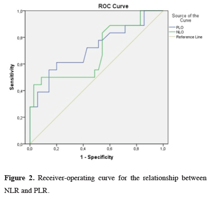
The cutoff NLR was obtained regarding the differences between the ISF and CSF groups by using ROC analysis. For the NLR, the AUC was estimated as 58.5% (95% CI, 51.34–65.41%), and the best cutoff NLR was 2.1 (sensitivity, 88.9%; specificity, 28%) (Table 4, Figure 2).
)
)
Table 4: Prediction performance of the NLR and PLR for complication
 Click Here to Zoom |
Figure 2: Receiver-operating curve for the relationship between NLR and PLR. |
The cut off PLR was obtained regarding the differences between the ISF and CSF groups by using ROC analysis. For the PLR, the AUC was measured as 70% (95% CI, 63.1-76.3%), and the best cutoff PLR was 123.8 (sensitivity, 72%; specificity 68%) (Table 4, Figure 2).
A multivariate logistic regression analysis using the laboratory parameters that have a significance level of p < 0.05 in the univariate analysis demonstrated that PLR (OR: 0.33; 95% CI: 0.1-1.06; p = 0.52), NLR (OR: 0.29; 95% CI: 0.1-1.06; p = 0.078), the difference between ISF and CSF groups was not statistically significant (Table 5).
)
Discussion
The mean platelet count, PLR, NLR, CKMB, LDH and duration of hospital stay were significantly higher in CSF group than in ISF group in our study results.
There is still debate whether the associated injuries affect the length of hospital stay or not. According to Wiener et al [16] the length of hospital stay varies with the presence of an accompanying pathology. Potaris et al [17] on the other hand reported that there is no significant difference in the length of hospital stay between SF patients with or without associated injuries. Perez et al [18] also reported that patients with associated injuries have longer hospital courses than ISF patients. In our study, average duration of hospitalization in sternal fractures was 4.5 ± 6.2. The average duration of hospitalization was significantly higher in CSF group than ISF group (p = 0.001).
In patients with SF, cardiac injury should be excluded. The incidence of cardiac injury following blunt chest trauma has been reported up to 90% [19]. And also, the frequency of myocardial contusion was reported as 18% in patients with SF [20]. In a recent study Perez reported that of 184 patients that underwent at least one of the three cardiac contusion work-up tests, 24.4% had abnormal ECG, 15.9% had abnormal Troponin, and 8.8% had abnormal ECHO findings [18]. All of our patients were monitored for ECG and cardiac enzyme levels. Patients with pathologic findings at ECG and elevated cardiac enzyme levels were evaluated with ECHO. We detected 17 (30.9%) ECG changes and 5 (13.5%) elevated cardiac enzymes in our patients. We also had three patients (9.3%) with minimal pericardial effusion and one them was in ISF group with a previous history of mitral regurgitation. Although, there was no statistically significant difference in the pres¬ence of abnormal ECG findings and cardiac enzyme eleva¬tion among patients in both groups in our study, higher levels of PLR in CSF group may indicate an undiagnosed cardiac injury.
The three most common causes of the fracture were motor vehicle accident 33 (60%), 20 fall (36.4%) and violence 2 (3.6%), respectively. Wojcik [20] reported that motor vehicle accidents (59%) were the most common cause of SF in their retrospective study. Rashid et al [21] reported that patient age ranged between 30 and 92 years and the mean age was 64. Velissaris et al [19] reviewed 73 patients with SF so that most of the cases were male (69.8%), with a median age of 51. In our study, the median age of the cases was 50 (19-90) years and the most frequent cause of the fracture was motor vehicle accident (60%).
NLR is a simple, cheap and effective biomarker that has been studied properly in the literature in order to predict severity of multiple diseases, including inflammatory conditions, neoplastic diseases like gastric cancer, thyroid cancer, breast cancer, and solid tumors [22,23]. But literature regarding their usage in trauma patients is scarce. According to our results, NLR were a useful indicator for severity of sternum fractures. In the present study, we found that the sensitivity, specificity, negative predictive value and positive predictive value of NLR count were 88.9, 28, 71.79, and 55.28%, respectively, with 58.5% diagnostic accuracy for a cut-off value of 2.1.
PLR has been identified and used for these purposes and as a sign of severe inflammation. In prior studies PLR were also investigated in patients with myocardial infarction, peripheral arterial occlusive disease, end-stage renal diseases, malignancies [15,24-27]. Lack of publication in the English literature regarding the relation between serum biomarkers and severity of SF didn’t allow the possibility of proper comparing our results, so that in our discussion we mainly presented our results about this topic. According to the results of our study, PLR were a useful indicator for severity of SF and the sensitivity, specificity, negative predictive value and positive predictive value of PLR count were 72, 68, 70.8, and 69.2%, respectively, with 70% diagnostic accuracy for a cut-off value of 123.8.
Our study had limited number of patients, which can be attributed to the rarity of sternal fractures and ISF being even rarer. Lack of publication in the English literature regarding the topic of the study didn’t allow the possibility of proper comparison of our results. The small patient population and the retrospective nature of the study do not allow us to draw any further conclusion.
To our knowledge, there is no literature data exists regarding relation between serum biomarkers and severity of SF. In our opinion, the present study is the first report about the association of NLR and PLR with the severity of SF. Even with the above-mentioned limitations, we can conclude that these results may indicate a clinically useful marker that can be easily and reliably measured from a blood sample to predict outcome of patients with SF. Further studies are necessary to externally cross-validate our findings in a larger cohort of SF patients.
Declaration of conflicting interests
The authors declared no conflicts of interest with respect to the authorship and/or publication of this article.
Funding
The authors received no financial support.