
Abstract
Introduction
In this case report we report the PET/CT findings a patient with GTS, and his successful treatment after a multidisciplinary planning.
Case Presentation
)
 Click Here to Zoom |
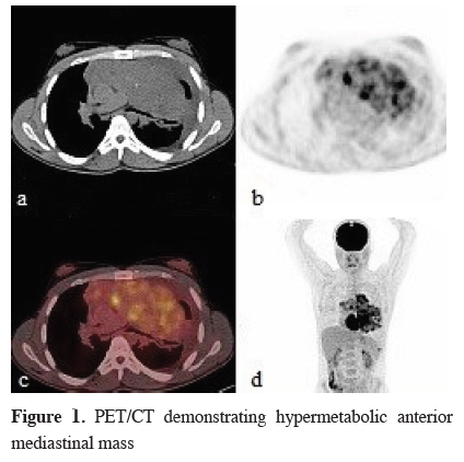
Figure 1: PET/CT demonstrating hypermetabolic anterior mediastinal mass |
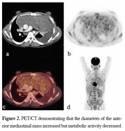
There was also a hypermetabolic nodule in the left pleural space and a non-metabolic left pleural effusion. Testicular ultrasound was normal. Serum AFP level was 3000 IU/dl (0-9 ng/mL), ß-hCG level was 555 IU/dl (0-2.67 mIU/mL). Pathologic diagnose was "mixed germ cell tumor" by CT guided tru-cut biopsy. The TNM classification was stage II. Three cycles of chemotherapy involving cisplatin, etoposide and bleomycin (BEP) therapy were applied. After the chemotherapy, patient was re-evaluated for treatment. PET/CT reported decreased metabolic activity (SUVmax: 5.5) but the tumor size increased to 21x9 cm (Figure 2).
)
 Click Here to Zoom |
Figure 2: PET/CT demonstrating that the diameters of the anterior mediastinal mass increased but metabolic activity decreased. |
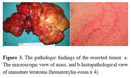
The AFP level was 14 ng/mL. The diagnosis of the overall examination was "Growing teratoma syndrome". Mediastinal mass was excised totally by median sternotomy. Macroscopic tumor size was 23 x 17 cm (Figure 3a). Pathological examination was reported as immature teratoma. Immature teratoma component originated from primitive mesoderm was 1-2%. Tumor included a necrotic area of %30-40 (Figure 3b).
)
 Click Here to Zoom |
Figure 3: The pathologic findings of the resected tumor. a-The macroscopic view of mass, and b-histopathological view of immature teratoma (hematoxylin-eosin x 4). |
Post-operative AFP level decreased to 9.3 ng/mL and ß-hCG remained within normal limits. After high-dose chemotherapy contains isophosphomide, carboplatin and etoposide, autologous stem cell transplantation (ASCT) was performed. The patient has been in the remission for two years.
Discussion
Determination of a mediastinal mass with radiological methods and elevation of serum AFP and ß-hCG may be enough for the diagnosis of NSGCT. Pathological examination provides an accurate diagnoses by revealing if an immature component exists. A tomography guided fine-needle biopsy or core biopsy can give a minimal invasive and successful diagnostic result. If this procedure is inadequate, it may be performed via a cervical mediastinoscopy, anterior mediastinotomy or thoracoscopy [4].
Mediastinal germ cell tumors frequently uptake FDG and PET/CT is used for staging and monitoring the metabolic response to chemotherapy, and also residue or recurrence of the tumor [5,6]. In this case the PET/CT was an essential tool for the diagnosis of GTS, showing a high metabolic activity and heterogeneity. After induction chemotherapy, the second PET/CT showed a decrease in the metabolic activity even though the diameters of the mass increased. The morphological and metabolic response mismatch findings supported the mixed nature of the mass [6].
The standard treatment of germ cell tumor must contain platinum-based regimen. The most common combination regimens are BEP (bleomycin etoposide, and cisplatin) and VIP (Isofosfamide, Etoposide, and Cisplatin) [7-9]. After three or four cycles of chemotherapy in case of full response, the patient must be followed without drugs. After chemotherapy, although tumor markers return to normal range, the mass grows up. In the presence of residual tumor or GTS surgical resection are proposed by most authors. [10-12] Sometimes the patient with GTS may refer in a bad condition and urgent operation may be necessary [10].
Sarkaria et al. [7] reported good prognostic factors in a series of 43% mixing type of totally 56 NSGCT patients as, R0 surgical resection after standard BEP chemotherapy, postoperative pathology demonstrates necrosis instead of teratoma, descending tumor marker levels after chemotherapy and prior stage I-II.
Walsh et al. [13] reported that an additional radical surgical resection to the initial standard or intensive chemotherapy after recurrence, increased the survey. Rodney et al. [14] suggested 3 poor prognostic factors as, serum ß-hCG level greater than 1,000 mIU/mL at diagnosis, presence of extra mediastinal disease and any mixed and single histologic diagnosis other than ectodermal sinus tumors. Accordingly in our case final pathological examination was reported as immature teratoma which is a prognostic factor.
Autologous stem cell transplantation following high-dose chemotherapy is defined as a treatment option for selected patients [8,15,16]. Bone marrow is affected by chemotherapy and regains the functions with ASCT. Bokemeyer et al [15] reported that according to "International Germ Cell Tumor Group Consensus Classification" the patients in the middle and poor prognosis groups benefit from high-dose chemotherapy and ASCT as a first treatment, instead of the standard treatment and the survey increases by %15. In this case, surgical excision followed by VIP chemotherapy and pathologic examination showed immature teratoma including 30%-40% necrosis. Although tumor markers and FDG uptake decreased the mass included more than 1% viable tumor cells so the high risk patient went to high-dose chemotherapy, and autologous ASCT.
In conclusion, we present this case as PET/CT findings of GTS was interesting, besides the patient was treated a successful instance of planning and treatment with medical oncology, thoracic surgery, hematology, and nuclear medicine clinics, i.e. multidisciplinary.
Declaration of conflicting interests
The author declared no conflicts of interest with respect to the authorship and/or publication of this article.
Funding
The author received no financial support for the research and/or authorship of this article.