
Abstract
Introduction
Case Presentation
)
 Click Here to Zoom |
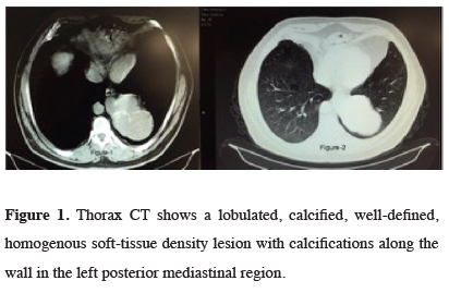
Figure 1: Thorax CT shows a lobulated, calcified, well-defined, homogenous soft-tissue density lesion with calcifications along the wall in the left posterior mediastinal region. |
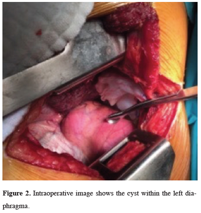
A left thoracotomy was performed for the resection of the lesion. Intraoperatively, a 6x8 cm sized cyst was found within the left hemidiaphragma. The mass has no connection with the left lung parenchyma. It was enucleated from the left hemidiaphragma (Figures 2,3). The diaphragma was repaired with a 6x8 cm sized polypropylene mesh. There were no intraoperative complications.
)
)
 Click Here to Zoom |
Figure 2: Intraoperative image shows the cyst within the left diaphragma. |
 Click Here to Zoom |
Figure 3: Excised cystic lesion. |
On gross examination, the cystic mass was multilocular, thin-walled and filled with red, yellow fluid. On microscopic sections, the cyst had smooth muscle tissue, cartilage and mucus glands on its wall. It was lined with ciliated cylindrical respiratory mucus epithelium, thus established a histopathologic diagnosis of intradiaphragmatic bronchogenic cyst.
Discussion
Bronchogenic cyst is the most common mass lesion in diaphragma in general population and the most common benign cystic process in infants. They are observed within congenital cysts [3,4]. Two-thirds of the patients are asymptomatic and the cyst is detected incidentally. Symptomatic patients generally present with chest pain, cough, dyspnea, fever and dysphagia. Chest pain is the most frequent symptom. In our case, chest pain complaint was observed as symptom and lesion was detected during radiographic examination.
It is often enough to apply plain chest radiography, thorax CT and bronchoscopy from among the laboratory methods for diagnosis. On plain film examinations, bronchogenic cysts present as spherical or oval masses with smooth outlines and soft tissue. CT is an excellent method for demonstrating the size, shape, position and margin characteristics of the cyst. It is also useful to show mass effect on adjacent structures, cyst attenuation, homogeneity and calcification. Barium swallow can be useful in dysphagic cases. In our case, radiology, especially CT, helped to a great extent about lesion’s nature, placement and size.
Life-threating complications such as airway obstruction, atelectasis, and compression are reported in pediatric age group. It is reported in current series that bronchogenic cysts in adults are more symptomatic. In our case, though it is adult, complications such as respiratory distress related with compression, atelectasis, and infection were not reported.
The cyst may show a solid appearance because of infection and its density increasing in parallel with its protein/calcium content. Cyst hydatid, lung abscess, infected air cyst, cavitary disease and malignancy should be kept in mind in differential diagnosis [2,5]. Bronchogenic cysts containing mucoid material are surrounded by a wall of bronchial cartilage, smooth muscle, elastic tissue and mucous gland [6,7]. In our case similar histopathological findings were reported.
Surgery may be considered as a treatment of choice even if the cyst is asymptomatic, to avoid complications such as infection, haemorrhage, or neoplasia within the cyst. Observation may be indicated for small, classic, asymptomatic cysts or high-risk patients.
Percutaneous catheter, drainage, sterile alcohol ablation or transbronchial cyst aspiration have been performed in selected cases [8,9]. Total excision of mediastinal bronchogenic cysts is adequate for treatment, minimal (wedge), segmental or lobar resection may be needed in parenchymal bronchogenic cysts [10].
Consequently, bronchogenic cysts must be kept in mind in all cystic lesions, in suppurated cyst appearance or in cases when tumor is not marginalized.
Declaration of conflicting interests
The authors declared no conflicts of interest with respect to the authorship and/or publication of this article.
Funding
The authors received no financial support.