
Abstract
Materials and Methods: Tracheae were collected from freshly slaughtered healthy sheep weighing 25-30 kg (2-3 years old) that were acquired from the local slaughterhouse. Twenty-eight tracheae were transported to the laboratory within an hour of slaughter and then dissected to remove the esophagus before obtaining 20-cm long tracheae. All tracheae were transected at the midpoint. In Groups 1 and 2, the transected tracheae were anastomosed by using interrupted 3-0 Vicryl sutures. In Groups 3 and 4, after anastomosing the transected tracheae with interrupted 3-0 Vicryl, the sites of anastomosis were reinforced by using Polyglycolic acid mesh. Groups 1 and 3 were tested for tensile strength, while Groups 2 and 4 were tested for pressure resistance.
Results: The median pressure was 49.00 ± 7.47 mmHg in the group anastomosed with only Vicryl, whereas it was measured as 70 ± 13.49 mmHg in the group on which the anastomoses were reinforced with PGA mesh. The median force needed to break anastomoses were 166.40 ± 24.28 Newton in the Vicryl-only group and 136.80 ± 34.93 newton in the PGA- reinforced group. The difference between the pressure resistance values of the Vicryl-only group and the PGA reinforced group was significant (p = 0.006). However, the difference with regard to the tensile strength was insignificant (p = 0.201).
Conclusion: We found that the reinforcement of the anastomosis site with polyglycolic acid mesh significantly increases the pressure resistance but does not significantly affect tensile strength.
Introduction
The most common complications associated with tracheal resection include dehiscence of the anastomosis, granulation tissue formation, and subcutaneous emphysema [2]. Complications that arise from tracheal surgery may result in re-stenosis, and therefore, it is important to prevent these complications. Many clinical and experimental studies have been conducted on reinforcement of the anastomosis. Polyglycolic acid (PGA) mesh is a biodegradable material which consists of PGA layers and oxidized cellulose mesh. Its long-term effects are not clear yet. Although PGA mesh has been used in the lung parenchyma to prevent air leakage, its application in tracheal surgery is not common [3,4].
We aim to investigate the effect of PGA mesh application on air leakage and tensile durability in tracheal resection and end-to-end anastomosis.
Materials and Methods
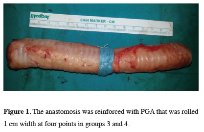
In Groups 1 and 2, the transected tracheae were re-anastomosed by using interrupted 3-0 Vicryl sutures (Medeks, Istanbul, Turkey), with a gap of 3-4 mm in between the sutures. In Groups 3 and 4, after re-anastomosing transected tracheae with interrupted 3-0 Vicryl, the sites of anastomosis were reinforced by using polyglycolic acid mesh (Neoveil, Gunze, Ayabe, Japan). The polyglycolic acid mesh, 1 cm in width, was wrapped all around the anastomosis and sutured with 3-0 Vicryl at four points of the anastomosis (Figure 1).
)
 Click Here to Zoom |
Figure 1: The anastomosis was reinforced with PGA that was rolled 1 cm width at four points in groups 3 and 4. |
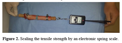
Groups 1 and 3 were subjected to tensile strength experiments and Groups 2 and 4 were subjected to pressure experiments. The tensile strength experiment was performed by using an electronic spring scale, with the force needed to break anastomosis being recorded [5] (Figure 2). For the pressure experiment, a sphygmomanometer was attached to a cuffed 8.5 mm endotracheal tube, with the pressure at air leakage observed being recorded [5] (Figure 3). SPSS for Windows (version17.0, IBM, Armonk, New York) was used for statistical analysis. All data are presented as the mean ± standard deviation. Comparison between group means was carried out using the Mann-Whitney U test at a significance level of p < 0.05
)
)
 Click Here to Zoom |
Figure 2: Scaling the tensile strength by an electronic spring scale. |
 Click Here to Zoom |
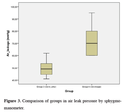
Figure 3: Comparison of groups in air leak pressure by sphygmomanometer. |
Results
)
 Click Here to Zoom |
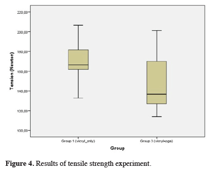
Figure 4: Results of tensile strength experiment. |
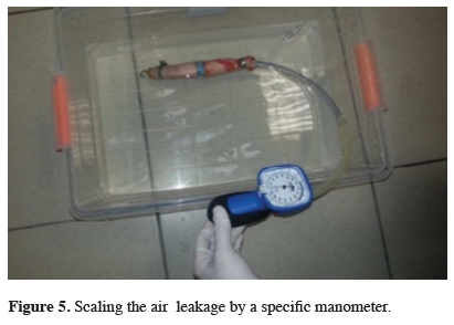
There was no significant difference between group 1 and group 3 with respect to the mean force required to break off the anastomoses (p = 0.201). In the pressure experiment, the mean pressure resulting in air leakage was 49.00 ± 7.47 mmHg (min 41mmHg-max 62 mmHg) in group 2 (Vicryl only) and 70.00 ± 13.49 mmHg (min 60 mmHg-max 95 mmHg) in group 4 (Vicryl + PGA) (Figure 5). The mean pressure required to cause air leakage was significantly higher in group 4 compared to group 2 (p = 0.006).
)
 Click Here to Zoom |
Figure 5: Scaling the air leakage by a specific manometer. |
Discussion
In tracheal anastomoses procedure, the surgical field is filled with saline to control air leakage after completion of the anastomosis. This control measure is done under 30-40 mmHg ventilation pressure when the cuff of the intubation tube is deflated and the patients mouth and nose are obstructed. Any air leakage detected must be repaired. The pediculated strap muscle flap can be used to reinforce the anastomosis line. Placement of a chin-to-chest suture for seven days postoperatively is also recommended to prevent extension of the neck. The site of anastomosis must be re-evaluated in cases of subcutaneous emphysema or air leakage through drainage catheter, which would either be a minimal dehiscence or a life-threatening separation of the anastomosis. Both must be repaired and a muscle flap must be used. [9]. The median pressure required to cause air leakage was 49.00 ± 7.47 mm Hg in the group in which the anastomosis was performed only with sutures, whereas it was 70 ± 13.49 mmHg in the group in which the anastomosis was performed with sutures and PGA mesh.
Polyglactin (Vicryl) and polydioxanone are commonly used suture materials for the different techniques of tracheal anastomosis. Primary anastomosis with interrupted 3/0 or 4/0 Vicryl sutures is the most preferred approach in tracheal stenosis [2,8,10]. After the resection, two traction sutures are usually placed on both sides of the proximal and distal trachea. Vicryl sutures are then placed one by one, leaving the knots outside the tracheal lumen. Proximal and distal traction sutures are used to feel the tension. There is no way to determine the acceptable tension other than the experience of the surgeon [1]. In our study, in order to preserve the cartilages, the tracheae were transected at the midpoint, through two tracheal rings. End-to-end anastomosis was performed by using interrupted 3/0 Vicryl sutures. PGA mesh is a bio-degradable material composed of polyglactic acid sheets and oxidized cellulose mesh. It is a synthetic, absorbable material which is used for tissue reinforcement.
Although its long-term effects are not clear, it is used in lung surgery to prevent parenchymal air leakage. However, until now, its effects on tracheal surgery have not been reported. PGA mesh is not only used in lung surgery but also in pancreas and spinal surgery as well to prevent leakage [3,4,11,12]. It is also used in liver surgery to prevent bile leakage [13]. To the best of our knowledge, there are no reports in the literature on the use of PGA mesh in tracheal surgery. Nevertheless, it has been reported that the use of PGA mesh in iatrogenic injuries of the distal trachea and main bronchus shows promising results [14]. PGA mesh is most commonly used in the lung parenchyma to prevent air leakage with or without fibrin glue. Its application in pneumothorax with Video Assisted Thoracic Surgery (VATS) has been reported to decrease complications like air-leakage and recurrence of pneumothorax. Its use on the site of the staple may prevent air-leakage, new bulla formation and recurrences [3]. As cautioned against in the study just cited, chemical pleurodesis and fibrin glue are not used in young patients with pneumothorax. Additional studies may help to better understand the effects of PGA mesh.
Air leakage is common complication of lung parenchymal surgery, and it is one of the most common causes of morbidity after lung resection [15,16]. Persistent air leakage for more than five days may result in atelectasis, pneumonia, and empyema [17]. Despite the use of different surgical techniques and materials to decrease air leakage, it still has a high incidence. It is observed in 48-70 % of the cases intraoperatively and may persist for more than seven days in 15-18% of the cases. There is still no ideal method for prevention [18]. The literature shows that PGA has been used with fibrin glue for pulmonary air leakage. Fibrin glue has been shown to be effective in the prevention of air leakage [19,20].
The reconstruction techniques applied for tracheal anastomoses after tracheal resection are similar to each other, but different tissues and materials are used to reinforce the anastomoses. For example, strep muscles, fibrin glue, bovine pericardium, Polytetrafluoroethylene (PTFE), and PGA+fibrin glue are some of the materials and tissues used [9,21].
In a previous ex-vivo study, it was reported that the application of n-butyl-2-cyanoacrylate to the site of the anastomosis in the trachea had beneficial effects in terms of intraluminal pressure. However, another study on rats revealed this application’s inflammatory effects, which may undermine its usage in vivo [5,22].
In conclusion, while different techniques to reinforce tracheal anastomoses are still under investigation, our study was nonetheless able to show that PGA mesh may be an effective method to reinforce tracheal anastomoses for the prevention of air-leakage. However, further clinical and experimental investigations are still needed for confirmation of these effects.
Declaration of conflicting interests
The authors declared no conflicts of interest with respect to the authorship and/or publication of this article.
Funding
The authors received no financial support.