
Abstract
Introduction
In this case report we present the successful treatment of a rare case of expectorated metastatic leimyosarcomas excised with rigid bronchoscopy and argon laser.
Case Presentation
)
 Click Here to Zoom |
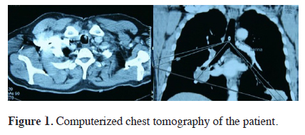
Figure 1: Computerized chest tomography of the patient. |
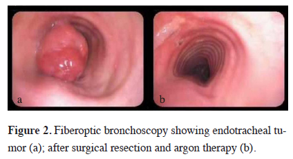
Fiberoptic bronchoscopy under local anesthesia revealed a large, white, glistening endotracheal lesion arising from the left anterolateral wall and obstructing %70 percent of the trachea (Figure 2a). With general anesthesia, a rigid bronchoscopy was performed and the lesion was successfully excised and argon coagulation was applied for hemostasis (Figure 2b). The specimen showed the identical pathology with the previous expectorated tissue. The patient had no complications, has recovered from is symptoms and was discharged from our clinic the next day. Two weeks after treatment, trachea was confirmed to be normal.
)
 Click Here to Zoom |
Figure 2: Fiberoptic bronchoscopy showing endotracheal tumor (a); after surgical resection and argon therapy (b). |
Discussion
As a conclusion, the patients who had leiomyosarcoma neoplasms should be evaluated for endotracheal metastasis in case of acute airway obstruction. These lesions could be resected safely by endotracheal approach.
Declaration of conflicting interests
The author declared no conflicts of interest with respect to the authorship and/or publication of this article.
Funding
The author received no financial support for the research and/or authorship of this article.