
Abstract
Introduction
Case Presentation
)
)
 Click Here to Zoom |
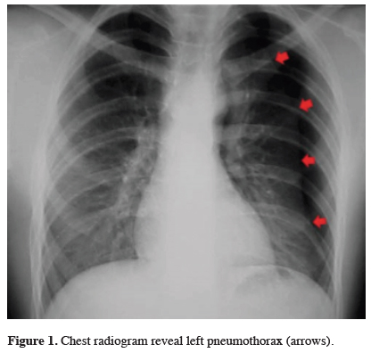
Figure 1: Chest radiogram reveal left pneumothorax (arrows). |
 Click Here to Zoom |
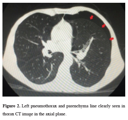
Figure 2: Left pneumothorax and parenchyma line clearly seen in thorax CT image in the axial plane. |
 Click Here to Zoom |
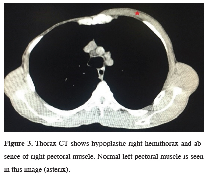
Figure 3: Thorax CT shows hypoplastic right hemithorax and absence of right pectoral muscle. Normal left pectoral muscle is seen in this image (asterix). |
 Click Here to Zoom |
Figure 4: One of the clinical component of Poland’s syndrome: hand abnormalities (absence of medial phalanges in right hand) Normal left medial phalanges are seen in this image (asterix). |
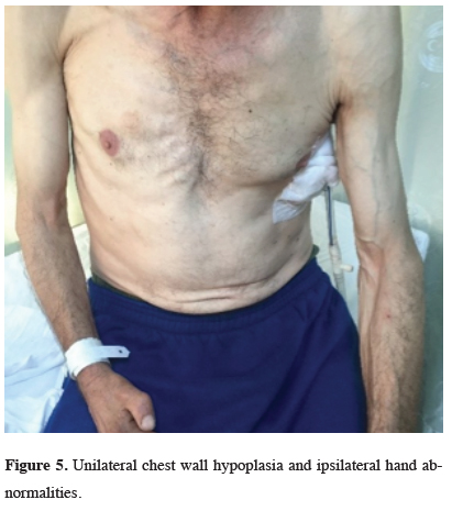
Tube thoracostomy was performed from the fifth intercostal space on midaxillary line (Figure 5). His symptoms relieved after the intrapleural air was evacuated.
)
 Click Here to Zoom |
Figure 5: Unilateral chest wall hypoplasia and ipsilateral hand abnormalities. |
Lung expansion was obtained on 4th day of drainage. Chest tube was removed and patient was discharged at 5th day of hospitalization.
Discussion
In literature, there are few reports about the familial occurrence of Poland’s syndrome but, it was thought to be an intrauterine pathology rather than a genetic defect [4]. The etiology is unknown but hypoxia during in the sixth to seventh week of gestation is thought to be a main etiological factor causing hypoplastic or aplastic musculoskeletal abnormalities. The possible cause of the hypoxia would be arterial spasm or an arterial malformation [4,5].
Poland’s anomaly with spontaneous pneumothorax has been previously reported only in few cases [6,7]. We detected only one case Poland’s anomaly with contralateral spontaneous pneumothorax.
In literature, emphysema has been shown to be associated with spontaneous pneumothorax and it is defined as an “abnormal permanent enlargement of air spaces distal to the terminal bronchioles”. Main pathology is the destruction of lung’s epithelial barrier causing a decrease in pulmonary elastic recoil. Cigarette smoke exposure is generally the main activator of destruction cascade. In addition, hypoxia would aggravate oxidative stress and protease production resulting emphysematous changes in the lung.
Considering the pathophysiology of Poland’s syndrome based on hypoxia, etiological factors that cause this syndrome may also be responsible for the occurrence of bulla formation and emphysematous changes in lung. In addition, different brachial arch blood supply defects might be responsible for the contralateral occurrence of pneumothorax [7].
Consequently a little is known about the relationship between pathogenesis of Poland’s syndrome and pneumothorax. More such should be presented to identify the relationship of these two diseases.
Declaration of conflicting interests
The authors declared no conflicts of interest with respect to the authorship and/or publication of this article.
Funding
The authors received no financial support.