






Summary
Plasmacytomas are localized proliferations of plasma cells in bone marrow. Extraosseous tissue involvement is uncommon. Herein, we report a case of mediastinal extramedullary plasmacytoma in a 54-year-old woman. A 85 mm lesion was reported in computed chest tomography of the patient who referred to ourvhospital with dyspnea. Since interventional radiologists did not think the patient was suitable for percutaneous biopsy, a surgical biopsy was performed and the pathology reported a plasmacytoma. Various investigations were done to rule out multiple myeloma. Since no result in favor of multiple myeloma was obtained, mass was diagnosed as extramedullary plasmacytoma. Radiotherapy was scheduled for the patient.Introduction
Malignant plasma cell dyscrasias occur due to the proliferation and differentiation of type B lymphocytes into autonomous, immunoglobulin producing plasmacytoid cells. Bence-Jones and Dalrymple first described it, in a patient with diffuse bone pain and proteinuria in 1846. Extramedullary plasmacytoma (EMP) is a plasma cell neoplasm of the soft tissue without having bone marrow involvement or other systemic features of multiple myeloma. EMP is a rare tumor and mostly detected in the upper aero-digestive tract. It is also very unusual for EMP to occur in mediastinum as a primary solitary lesion [1-5]. Herein we report an extremely unusual presentation of an EMP arising from the posterior mediastinum.Case Presentation
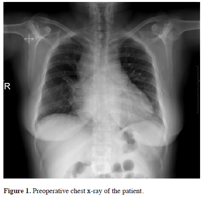
A 54-year-old woman referred to our clinic with progressive dyspnea. Her surgical history includes appendectomy, cesarean section and a lumbar hernia operation. Her physical examination was considered normal except a lack of ventilation in the right hemithorax. No goiter, hepatosplenomegaly or significant lymphadenopathy was noticed. Laboratory blood tests revealed haemoglobin level 11.9 gr/dL, leukocyte level 3.2 × 10³/μL (neutrophils 53%, lymphocytes 35%, eosinophils and basophils both 0%) and thrombocyte level 136 × 103/μL. Her renal and liver functions were reported normal. The plain chest X-ray revealed a smooth, mass-like lesion in the right paratracheal region (Figure 1).)
 Click Here to Zoom |
Figure 1: Preoperative chest x-ray of the patient. |
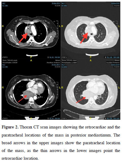
Chest computerized tomography (CT) revealed a mass, which was spreading to the subcarinal and right paraesophageal regions. The size of the mass was 49×48 and 84×58 mm in the right paratracheal and retrocardiac areas, respectively (Figure 2).
)
 Click Here to Zoom |
Figure 2: Thorax CT scan images showing the retrocardiac and the paratracheal locations of the mass in posterior mediastinum. The broad arrows in the upper images show the paratracheal location of the mass, as the thin arrows in the lower images point the retrocardiac location. |
Positron Emission Tomography (PET) is performed and the SUVMax of the mass is found to be 2.9. No pathologic FDG involvement is reported for any other tissue. As transthoracic needle sampling was not recommended by the interventional radiology department, surgical biopsy of the mass was planned. Instead of minimally invasive methods such as EBUS or EUS, surgical procedures were considered convenient also due to the demand of the oncologist for a larger piece of specimen for further investigations. Since, single lung ventilation was not considered to be convenient by the anesthesiologists due to mass’ pressure on trachea, thoracoscopic surgery options were called off and a 10 cm long, right limited thoracotomy was performed. A 10 cm mass was observed in the posterior mediastinum between the carina and esophagus. Due to the localization of the mass, its relation with the surrounding structures and the bleeding during the operation, complete resection couldn’t be performed. Multiple biopsies were taken for pathological investigation and the thoracotomy incision was closed routinely. The pathologic examination of the mass was reported as plasmocytoma with lambda light chain restriction. CD138, MUM-1 and lambda light chain immunohistochemical studies were reported positive while, CD3, CD68, CD34, CD20, kappa light chain and mast cell triptase were negative. The patient was consulted to the oncology department. During the evaluation in the oncology department her bone survey was studied. Neither focal litic bone lesions, which might have been related to multiple myeloma, nor any vertebral collapse was noticed. Bone marrow biopsy was performed and revealed no pathologic pattern of cell distribution. Both serum and urine protein electrophoresis were performed and gama peaks sighted. No M-protein or urine Bence-Jones protein was detected. Consequently, the tumor was diagnosed as a plasmacytoma and radiotherapy was scheduled.
Discussion
Extramedullary plasmacytomas (EMP) constitute approximately 3% of the plasma cell neoplasms. It is mostly seen around the 6th decade of life and 3-4 times more in men with respect to women [4]. EMP diagnosis is verified only if there is no evidence of multiple myeloma. To rule out multiple myeloma, there should be no histopathological evidence of plasma cell dyscrasia in bone marrow, with no clinical symptom such as bone pain, renal failure or anemia and in addition no presence of focal lytic bone lesions, which were all confirmed by CT, bone survey, bone marrow aspiration investigation and blood tests in our case [2,6]. Plasma cell neoplasms are considered more sensitive to radiotherapy with respect to other types of treatment. Mostly surgical resection of the tumor is combined with RT. Most of the patients are cured after surgery and RT. Local recurrence is shown in nearly 30% of the patients. In 40% of the patients, multiple myeloma emerges. An increase in the baseline level of serum monoclonal proteins or detection of Bence-Jones protein in urine may signify the recurrence of the disease or the onset of multiple myeloma. Dissemination into multiple myeloma has been reported in 10-30% of the patients with EMP in the first 2 years [3,7]. Thus, long-term follow-up is mandatory in these rare tumors.
Declaration of conflicting interests
The authors declared no conflicts of interest with respect to the authorship and/or publication of this article.
Funding
The authors received no financial support.
Reference
1) Lee SY, Kim JH, Shin JS, Shin C, In KH, Kang KH, Se Yoo SH. A case of extramedullary plasmacytoma arising from the posterior mediastinum. Korean J Intern Med 2005; 20: 173-6.
2) Shukla A, Bansal V, Bhutani R, Kumar G, Sharma J, Solanki J, Mehta A. Extramedullary plasmacytoma presenting as a mediastinal mass. Turk J Hematol 2011; 28: 228-31.
3) Luh SP, Lai YS, Tsai CH, Tsao TC. Extramedullary plasmacytoma (EMP): Report of a case manifested as a mediastinal mass and multiple pulmonary nodules and review of literature. World J Surg Oncol 2007; 5: 123.
4) Miyazaki T, Kohno S, Sakamoto A, Komori K, Sasagawa I, Futagawa S, Hara K. A rare case of extramedullary plasmacytoma in the mediastinum. Intern Med 1992; 31: 1363-5.
5) Soesan M, Paccagnella A, Chiarion-Sileni V, Salvagno L, Fornasiero A,Sotti G, et al. Extramedullary plasmacytoma: clinical behaviour and response to treatment. Ann Oncol 1992; 3: 51-7.