
Abstract
Introduction
Lipomas, one of the most common benign soft tissue lesions, may mimic malignant masses with their clinical and radiological manifestations. They rarely exhibit excessive growth and can be termed as giant lipoma, if they are greater than 10 cm in diameter. With their slow growth, well-encapsulated and mobile characteristics, lipomas can be differentiated from other tumoral formations based on clinical findings, and they may be present in the breast tissue.
Case Presentation
)
 Click Here to Zoom |
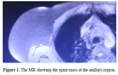
Figure 1: The MR showing the giant mass at the axillary region. |
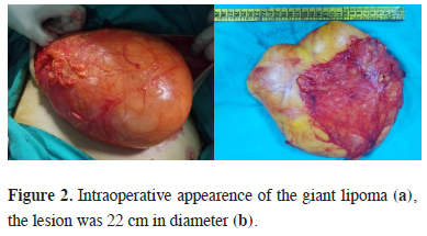
Since the possibility of malignancy could not be eliminated on the examinations carried out, surgical intervention was planned for diagnosis and treatment. The patient was placed in the right posterolateral position and via a 10 cm incision across the right anterior axillary and nipple line, which was above the fifth costa, the mass was observed under latissimus dorsi muscle, and excised totally. No costal invasion was present (Figure 2). Histolopathologic examination confirmed lipoma.
)
 Click Here to Zoom |
Figure 2: Intraoperative appearence of the giant lipoma (a), the lesion was 22 cm in diameter (b). |
The patient was discharged on the third postoperative day uneventfully.
Discussion
In conclusion, lipomas, which are most commonly benign soft tissue lesions of the chest wall, can mimic malignant masses with clinical and radiological manifestations. Surgical interventions and pathological examinations performed for definitive diagnosis may bring surprising results. Especially in cases of ignored pain and swelling, necessary diagnostic procedures should be performed promptly, and large resection should not be avoided in operable cases.
Declaration of conflicting interests
The authors declared no conflicts of interest with respect to the authorship and/or publication of this article.
Funding
The authors received no financial support