
Abstract
Introduction
Case Presentation
)
 Click Here to Zoom |
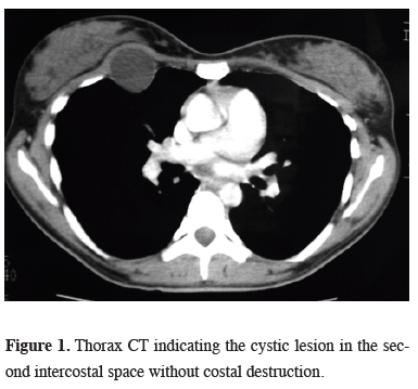
Figure 1: Thorax CT indicating the cystic lesion in the second intercostal space without costal destruction. |
Surgical excision was planned for the exact diagnosis and the treatment. A thoracic incision was made through the third intercostal space, and after dissection of the subcutaneous tissue and pectoral muscles, a cystic lesion of 4x4 cm diameter was visualized between the muscles of intercostal region that was fluctuating on palpation. The ribs were not invaded and the cystic lesion was bulging through the intrapleural space. It was totally excised with blunt and sharp dissection. Cuticular membrane of cystic lesion was confirmed on the specimen. There was no complication postoperatively. Histopathologic examination confirmed that the lesion was hydatid cyst. Albendazole was administered after the operation. The patient was discharged on day 5 uneventfully.
Discussion
To distinguish of chest wall hydatid cyst from other masses is difficult, as primary chest wall involvement of hydatid disease cannot be considered first by physicians. Chest radiographs usually show us unruptured cysts as round homogeneous well-defined densities but like in the patient reported by Demir et al. the disease can destruct the ribs and can mimic a chest wall tumor [7]. Also CT is not pathognomonic in these patients.
In patients with a history of surgery for lung hydatid cyst, masses of the thoracic wall must be considered as the spread the disease to the chest wall [5]. The pulmonary cysts may rupture spontaneously or during surgery. Rupture of the pulmonary hydatid cysts into the pleural space may contaminate the adjacent organs and this may result to development of pleural or chest wall hydatid cysts. Likewise, in patients with concomitant cystic lesions especially in the lung and liver with the cystic chest wall masses may be considered as clues of the chest wall hydatid cyst. Although in our patient we did not detect a clue considering hydatid cyst, we decided surgery because it would be appropriate for the diagnosis and treatment of this type of cystic lesions of the thoracic wall. In our opinion, percutaneous biopsy is not suitable in such patients due to the risk of rupture of the cyst and dissemination of the infection.
In this case, the intercostal involvement is via systemic circulation i.e. to right heart after passing the common sites, liver and lung. The involvement of body’s unexpected portions can be seen rarely with systemic circulation of hydatid cyst [1].
Surgical treatment of the disease is radical removal of involved chest wall. In this case, only the intercostal muscle was involved, therefore the cyst and muscle around it were totally excised. Medical treatment should be used after surgery to avoid recurrences. Albendazole treatment after surgery has a high response rate without recurrence [8]. There are studies that albendazole should be used preoperatively [9]. Although preoperative albendazole treatment can sterilize cyst cavity, it can also cause rupture of the intact cyst and may cause further complications.
In conclusion, hydatid cyst must be considered in the differential diagnosis of chest wall masses when dealing with chest wall tumors even without apparent risk factors. Appropriate treatment of primary chest wall hydatid cyst is surgical excision.
Declaration of conflicting interests
The authors declared no conflicts of interest with respect to the authorship and/or publication of this article.
Funding
The authors received no financial support.