
Abstract
Introduction
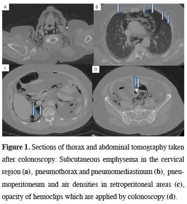
In this report, we present a patient with bilateral pneumothorax, pneumomediastinum, subcutaneous emphysema, pneumoperitoneum due to iatrogenic colon perforation during diagnostic colonoscopy. Unfortunately, during the treatment of pneumothorax, an iatrogenic hemothorax also developed all iatrogenic injuries were treated successfully.
Case Presentation
)
 Click Here to Zoom |
Figure 1: Sections of thorax and abdominal tomography taken after colonoscopy. Subcutaneous emphysema in the cervical region (a), pneumothorax and pneumomediastinum (b), pneumoperitoneum and air densities in retroperitoneal areas (c), opacity of hemoclips which are applied by colonoscopy (d). |
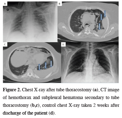
24F apical tube thoracostomy was performed under local anesthesia for treatment of left sided pneumothorax. The control chest X-ray showed complete expansion of the left lung (Figure 2). Antibiotic treatment was arranged and oxygen therapy was administered by for pneumomediastinum, pneumoperitoneum and subcutaneous emphysema. On the second postoperative day, pleural effusion and subpleural hematoma was seen on the chest radiography and a 32F thorax catheter was inserted to left hemithorax and approximately 500 cc of blood was drained (Figure 2). The patient was followed up on daily chest radiographs, hemogram and C- reactive protein levels.
)
 Click Here to Zoom |
Figure 2: Chest X-ray after tube thoracostomy (a), CT image of hemothorax and subpleural hematoma secondary to tube thoracostomy (b,c), control chest X-ray taken 2 weeks after discharge of the patient (d). |
The patient was followed by general surgeons and gastroenterologists on daily bases. Abdominal examination was normal and oral intake was allowed on the 4th day. Apical thorax catheter was terminated on the 3rd day and basal thorax catheter was terminated on the postoperative 5th day. The patient was discharged uneventfully. 6th month follow up of the patient as normal. A written consent form was obtained from the patient for this scientific publication.
Discussion
Colonoscopic perforation can occur by various mechanisms; direct mechanical trauma, thermal injury or barotrauma (because of massive air insufflation) [5,6]. Many factors such as old age, female sex, previous abdominal-pelvic surgery, adhesions, diverticulosis, severe colitis, inflammatory bowel disease, malignancy, radiation therapy, low albumin level etc. have been identified for increased risk [1-3,6,7]. Unfortunately, our patient had three risk factor together as; advanced age, colon diverticulum and female sex.
Perforations may be located in the intraperitoneal/extraperitoneal part of the colon, or occur with combination, although clinical signs and symptoms variable according to perforation location [1-3]. When perforation develop in the intraperitoneal part of colon, abdominal complaints may be the first symptoms such as pain, distension or rigidity but, if the perforation is in extraperitoneal part of the colon or rectum, atypical symptoms may develop such as dyspnea, tachypnea and subcutaneous emphysema [1,2,5].
The pathophysiological mechanism of subcutaneous emphysema and pneumomediastinum might be explained due to the presence of a visceral space, which goes from the neck through the mediastinum and retroperitoneum [4-8]. This visceral space follows the trachea and esophagus along the chest and creates a tract for air. The space continues inferiorly through the diaphragmatic hiatus and reaches to the retroperitoneum [4-8]. When air enters the retroperitoneum, it rises by pressure effect along the fascial planes, follows the large vessels, progress through the diaphragmatic hiatus, and reaches to the mediastinum and subcutaneous neck tissues [4-10]. Large amount of air may leak from the mediastinal pleura to the pleural cavity [5,8,9]. Alternatively, free air in the peritoneal cavity may permeate through to intrapleural cavity with small diaphragmatic defects or fenestrations. Air may spread to many places with similar mechanisms, and even cases with atypical findings such as pneumoscrotum have been reported [11].
Clinical suspicion, physical examination and radiologic findings are important in diagnosis. Accompanied by clinical findings, it has been reported that sub-diaphragmatic air in abdominal radiography is an important sign and can be diagnosed in approximately 87% of cases [2]. Chest radiography may also be helpful in cases who present with pneumothorax [3-7]. Abdominal computed tomography may provide information about the location of perforation [1-5].
Early diagnosis of colon perforation is an important issue and it also affects the choice of treatment likelihood; nonoperative management, primary repair, intestinal resection with a primary anastomosis or double stage procedure [2]. In only a rare portion of patients may diagnosed during the procedure like in our case. Corey W et al [2] examined 180 patients who developed colon perforation after colonoscopy and reported 140 of patients were diagnosed within 24 hours. Only 42 patients were diagnosed during colonoscopy. Peritonitis findings may progress and surgical treatment may become more difficult for general surgeons in patients with late diagnosis [2]. It has been reported most of the cases have to undergone surgical treatment and endoscopic repair can be performed in a very limited number of cases in the literature [1,2]. In this case, colonoscopy was performed by an experienced gastroenterologist, perforation was noticed during colonoscopy and repaired with endoscopic hemoclips. The patient was carefully followed up by the general surgery clinic after the procedure and there were no abdominal complaints.
For the treatment of pneumomediastinum and subcutaneous emphysema oxygen inhalation and supportive therapy were preferred in this patient. Since the etiology of pneumomediastinum was known and tracheobronchial injury was not suspected, additional diagnostic procedures such as bronchoscopy were not performed. For bilateral pneumothorax treatment, left tube thoracostomy was used and the patient was followed up with chest radiographs. Unfortunately, an iatrogenic hemothorax also developed in our patient secondary to tube thoracostomy. Therefore, a second tube thoracostomy was performed and the patient was treated without any additional problems.
In conclusion, follow-up period after colonoscopy is an important issue. Abnormal abdominal examination findings such as pain, distention, rebound, rigidity etc. are well known by gastroenterologists and general surgeons. Atypical findings such as subcutaneous emphysema, dyspnea, hoarseness, impaired voice quality may be seen in a small minority of patients. When these atypical findings are encountered, differential diagnosis such as pneumothorax, pneumomediastinum should be kept mind as a sign of iatrogenic colon injury. Cooperation with gastroenterologist, thoracic surgery and general surgery clinics will be effective in achieving successful results.
Declaration of conflicting interests
The authors declared no conflicts of interest with respect to the authorship and/or publication of this article.
Funding
The authors received no financial support.