
Abstract
Introduction
We here present an incidentally diagnosed giant cystic lymphangioma, which was located in the right paratracheal area and excised via video-assisted thoracoscopic surgery (VATS).
Case Presentation
)
 Click Here to Zoom |
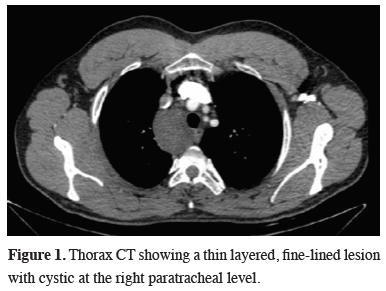
Figure 1: Thorax CT showing a thin layered, fine-lined lesion with cystic at the right paratracheal level. |
The physical examination was normal. He described a decrease in his exercise capacity during the last 3 years, without causing a problem to his daily routines. There were no abnormalities in the routine blood tests, EKG and pulmonary function test (FEV1: 4.61 L, %105.8, FVC: 4.62, %89.8)
Surgery was planned with the initial diagnosis of a bronchogenic cyst and considering the deep mediastinal localization, vascular surroundings and the abundance of cystic nature, no preoperative invasive diagnostic procedure was done.
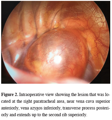
The exploration was done via two-port VATS, under general anesthesia with double-lumen endotracheal tube intubation. An uninoculated cystic mass, which was located in the right paratracheal area, in close relation to vena cava superior anteriorly, vena azygos inferiorly, transverse process posteriorly and extending up to second rib level superiorly was observed (Figure 2). After excision of the mediastinal pleura, the deep extension of the cyst between vena azygos, vena cava superior, and the brachiocephalic vein was seen. The cyst was aspirated which was clear and straw-like colored, to reach the root for complete dissection to prevent injury to vessels. After the root of the lesion was released from the posterior side of superior vena cava, it was separated from the mediastinal fatty tissue by using an endolinear stapler. After inserting one apicobasal chest tube, the operation was terminated without any complication.
)
 Click Here to Zoom |
Figure 2: Intraoperative view showing the lesion that was located at the right paratracheal area, near vena cava superior anteriorly, vena azygos inferiorly, transverse process posteriorly and extends up to the second rib superiorly. |
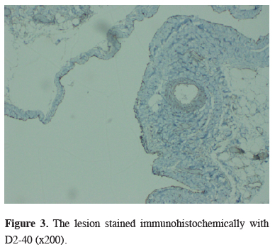
The patient was uneventfully discharged on postoperative day 3. In the histopathological examination of the lesion, CD31 and D2-40 antibodies were stained positive which was compatible with cystic lymphangioma (Figure 3).
)
 Click Here to Zoom |
Figure 3: The lesion stained immunohistochemically with D2-40 (x200). |
Physical examination was normal at three month follow-ups, and CT scan revealed no recurrences at the first-year control.
Discussion
Mediastinal cystic lymphangiomas are rarely seen lesions that are typically located in the superior mediastinum and at the right hemithorax. Bronchogenic cysts, thymic cysts or thymomas, coelomic cysts, enteric cysts, and ductus thoracicus cysts should be considered at the differential diagnosis. Most of the lymphangiomas detected in adulthood are asymptomatic. However, patients can be presented with symptoms such as chest pain, cough, dyspnea, stridor, dysphagia due to compression of intrathoracic structures. Hoarseness due to vocal cord paralysis and swelling of the neck due to venous compression may be observed.
The recommended radiological imaging methods for the diagnosis of the cystic lesion are x-ray and thorax CT. In our case, the chest x-ray showed a mediastinal widening and the thorax CT imaging revealed a cystic, well-circumscribed and homogenous soft tissue lesion. Three-dimensional ultrasound imaging is also useful for detecting fine septations compatible with lymphangioma. MRI scanning helps to accurately predict relations between the lymphatic chain and other mediastinal organs [1]. Cystic lymphangiomas characteristically immunohistochemically stained positive with CD31 and D2-40 [13].
The standard treatment modality is complete surgical excision via thoracotomy or VATS, as performed in our case. Surgical resection is necessary both to confirm the diagnosis and to prevent complications that may occur due to the compression effect on vital organs. Surgical removal of the cyst wall should be achieved completely. The success of totally excised cases is quite high. As we have performed, cysts in larger sizes can be completely resected after aspirating its content, to reveal the borders in the full sense and not to injure vascular structures.
Some authors predicted that cystic lesions might be in communication with the ductus thoracicus. Thus, in the preoperative period, the administration of olive oil from a nasogastric catheter was suggested. However, we did not prefer this method in this particular case [2].
Some authors suggest that these cysts can be excised by enucleation. Whichever the method is, it should be kept in mind that, there is a risk of recurrence in incomplete resections. We have not found any sign of recurrence after a one-year follow-up in our patient. Esme et al. reported significantly lower morbidity, mortality, and recurrence rate in patients undergoing surgery for mediastinal cysts, compared to conservative approaches [14]. Nowadays, surgical excision with VATS is used safely for curative treatment of many mediastinal cystic lesions in experienced centers. In the study of Ulaş et al. [15], VATS and conventional thoracotomy methods were compared in the surgical approach to mediastinal cysts, and it was statistically significant that the patients of the VATS group had lower hospitalization length and operation duration. Moreover, VATS patients had no increase in the means of intraoperative bleeding, postoperative complications, and incomplete resection risk. In that vein, we did not encounter any kind of intraoperative or postoperative complications, and the patient was discharged from the hospital three days after surgery with complete recovery. In our center, the first choice for the approach to mediastinal cystic lesions has been excisional intervention with VATS.
In conclusion, the surgical approach is the curative treatment method for mediastinal cystic lymphangioma cases, and nowadays, the VATS procedure should be preferred primarily as it significantly reduces the operation time and postoperative hospital stay.
Declaration of conflicting interests
The authors declared no conflicts of interest with respect to the authorship and/or publication of this article.
Funding
The authors received no financial support.