
Abstract
Introduction
Case Presentation
)
 Click Here to Zoom |
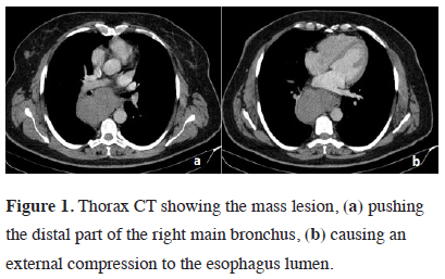
Figure 1: Thorax CT showing the mass lesion, (a) pushing the distal part of the right main bronchus, (b) causing an external compression to the esophagus lumen. |
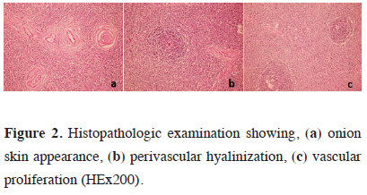
The right posterolateral thoracotomy incision revealed a mass lesion of approximately 14 cm located in the posterior mediastinum extending into the subcarinal area and azygos vein. The lesion was resected with the capsule. Invasion into the surrounding tissues was not observed. Final pathology was reported as Castleman’s disease (Figure 2). All symptoms of the patient, particularly dysphagia, regressed. The patient is still being followed up uneventfully in the 26th month.
)
 Click Here to Zoom |
Figure 2: Histopathologic examination showing, (a) onion skin appearance, (b) perivascular hyalinization, (c) vascular proliferation (HEx200). |
Discussion
It has two types as multicentric and unicentric [3,7]. Unicentric type is often asymptomatic, more common in women than in men, and no etiological factor has been identified [4,8,13]. It is seen in the 3rd or 4th decade of life and malignancy potential is low. It is mostly located in mediastinal lymph nodes. It may also be observed in the neck, axilla, retroperitoneal region, mesentery and pelvis [14]. Surgical treatment is curative and the symptoms regress after surgery [15,16]. Five-year survival has been reported to be 100% [2]. Our case was also diagnosed in the fourth decade, being consistent with the literature, and her symptoms improved after surgery. No etiological factor was identified. The lymph node was completely resected with the capsule and there was no invasion outwards.
Multicentric type is more common than unicentric and it is associated with HIV and HHV-8 more frequently [4,8,17]. Fever, night sweats, weakness and massive lymphadenopathy are the most common symptoms [18]. Surgical excision is the best method for diagnosis. However, it is not always possible to excise due to invasion into the surrounding tissue and hypervascularization. Systemic chemotherapy, antiretroviral agents, interferon alpha, immunomodulatory drugs, rituximab and corticosteroids are recommended for treatment [4]. Multicentric type has a worse prognosis than unicentric type [7]. Patients should be followed closely.
In conclusion, Castleman’s disease is a rare lymphoproliferative disease. It is mostly located in the mediastinum. Surgical excision is curative for localized type, dramatically improving the symptoms and the prognosis is excellent.
Declaration of conflicting interests
The authors declared no conflicts of interest with respect to the authorship and/or publication of this article.
Funding
The authors received no financial support.