
Abstract
Introduction
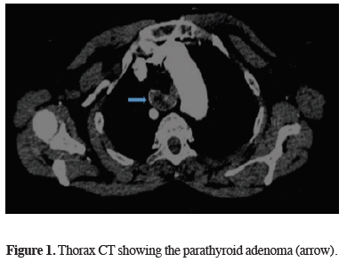
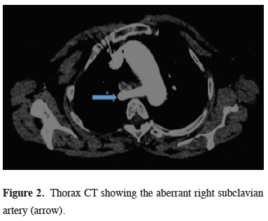
Here, we aimed to present a patient with the coexistence of mediastinal parathyroid adenoma and aberrant right subclavian artery, which is the most common intrathoracic major arterial anomaly.
Case Presentation
)
)
 Click Here to Zoom |
Figure 1: Thorax CT showing the parathyroid adenoma (arrow). |
 Click Here to Zoom |
Figure 2: Thorax CT showing the aberrant right subclavian artery (arrow). |
PTH level was 1225 pg/mL , serum calcium level was 11.4 mg/dL, and serum thyroid-stimulating hormone (TSH) value was 6.56 mIU/L. (The normal values for PTH, calcium and TSH is 15-65 pg/mL, 8.5- 10.5 mg/dL and 0.4-4 mIU/L respectively).
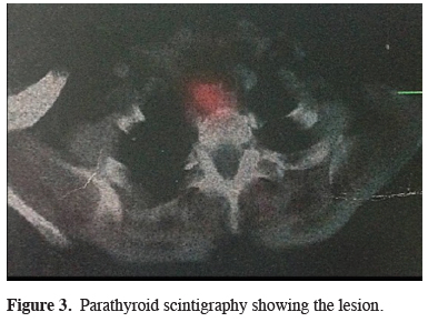
Parathyroid scintigraphy was performed with technetium 99, and the mass was evaluated in favor of parathyroid adenoma (Figure 3).
)
 Click Here to Zoom |
Figure 3: Parathyroid scintigraphy showing the lesion. |
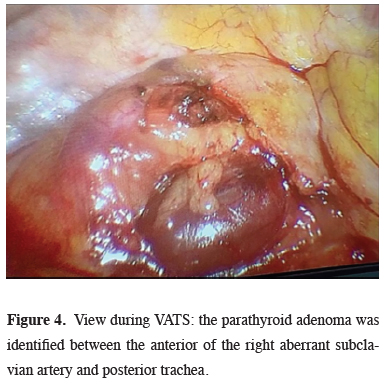
Based on these findings, mediastinal mass excision was performed via right videothoracoscopic approach. A utility port of 3 cm was placed at the fourth intercostal space at the anterior axillary line. One additional 1.5 cm port was then placed at the midaxillary line. After exploring the mediastinum, the parathyroid adenoma was identified between the anterior of the right aberrant subclavian artery and posterior trachea. Using a harmonic scalpel, the parietal pleura was dissected while carefully avoiding the ARSA (Figure 4).
)
 Click Here to Zoom |
Figure 4: View during VATS: the parathyroid adenoma was identified between the anterior of the right aberrant subclavian artery and posterior trachea. |
The parathyroid adenoma was then dissected free from the surrounding tissue. At the histopathological examination, the lesion with lobular appearance, dirty - yellow color and bleeding, was reported to be compatible with parathyroid adenoma. PTH level was 53.16 pg / mL and serum calcium level was 8.9 mg / dL on the first of postoperative day. The patient was discharged uneventfully at postoperative day 4.
Discussion
It should be kept in mind that hyperparathyroidism may be a component of multiple endocrine neoplasia type 1 (MEN), pituitary tumor, and pancreatic islet tumor [2]. Although it is mostly located in the parathyroid gland, approximately 25% of the patients have an ectopic location [1]. Thymus (38%), retro / paraesophageal space (31%), thyroid (18%), mediastinum (6%), and carotid sheath (3%) are the most common sites of ectopic localization [3]. Ectopic inferior parathyroid glands are usually located in the anterior mediastinum within the thymus, while superior parathyroid glands are located in the posterosuperior mediastinum. Since the size is generally small, it is crucial to use preoperative technetium 99 (99mTc) scintigraphy for the localization.
Aberrant right subclavian artery (ARSA) or Lusoria artery is the most common vascular anomaly of the aortic arch with a rate of 0.5-2.5% [4]. It is a rare anatomical variation in which ARSA originates directly from the distal part of the aortic arch instead of brachiocephalic artery. Although it is mostly localized in the retroesophageal site, it may be located in anterior of the trachea or between the esophagus and trachea. The majority of cases with ARSA are asymptomatic. Retroesophageal form is the most commonly seen form that patients may complain of dysphagia due to compression of the esophagus. Anterior tracheal localization may be the cause of dyspnea. Surgery for ASA is offered in just case of being symptomatic.
99TC-MIBI scintigraphy is not a definite method but has a sensitivity of 80-90% [5]. False positivity may be detected in patients with lung cancer metastasis or patients with mediastinal tumors such as thymoma, seminoma, and lymphoma. False negativity may be seen in patients with multiple adenomas or hyperplasia [6]. Also, thoracic computed tomography (CT) and magnetic resonance imaging are helpful to determine the anatomic localization and the relationship of mediastinal parathyroid adenoma with the other structures.
CT can detect lesions over 1.5 cm in size. Although reviews are reporting that intraoperative gamma probes can be used for small-sized lesions [7], some surgeons suggest that this method will not be beneficial due to large vessels and heart activities [8]. Intravenous methylene blue is another option to localize ectopic glands. In some cases, intraoperative parathyroid hormone assay is used to be sure that the excised mass is a parathyroid adenoma or not.
The definitive treatment of parathyroid adenoma is surgery. Surgical methods such as cervical approach, video-assisted mediastinoscopy, sternotomy, thoracotomy, video thoracoscopy should be chosen according to the location of the mass. The cervical approach is the preferred method for adenomas located at or above the aortic arch level. Thoracotomy, sternotomy, mediastinotomy, or video thoracoscopy (VATS) is more appropriate for lesions located under the aortic arch or deeper in the mediastinum [9]. Parathyroid adenoma excision using video thoracoscopy was first reported in 1994 by Prinz et al. Subsequently, it is reported that VATS is a feasible and safe approach for mediastinal parathyroid adenoma [10]. Since then, video thoracoscopy has been used more widely. VATS offers more advantages over both thoracotomy and sternotomy. It allows better visualization of the tumor and exposure of the mediastinum, excellent access, shorter operative time, shorter tube dwell time, shorter hospital stay and reduced postoperative pain. Also it is superior in the cosmetic way when compared to sternotomy and thoracotomy scars [11]. Complications such as pleural effusion, hematoma, pneumothorax, wound site infection, mediastinitis, and sternal dehiscence are more likely to be seen in conventional methods like sternotomy and thoracotomy.
In conclusion, videothoracoscopic resection of mediastinal masses can be performed safely even in patients with aberrant vascular structure.
Declaration of conflicting interests
The authors declared no conflicts of interest with respect to the authorship and/or publication of this article.
Funding
The authors received no financial support.