
Abstract
Materials and Methods: Fifty-eight patients who were admitted to the Thoracic Surgery Outpatient Clinic in our center due to minor thoracic trauma between October 2017 and July 2018 were included in the study. All patients underwent chest radiography, and thorax US after a detailed history and physical examination were performed.
Results: Of the 58 patients included in the study, 33 (56.9%) were male, and 25 (43.1%) were female, while the mean age was 46.79 ± 15.75 years. While the rib fractures were detected in 24 patients with chest x-ray, US detected rib fractures in 47 patients. The comparison between the two examinations, the superiority of the US in the diagnosis of rib fracture was statistically significant compared to the chest radiography (p < 0,001).
Conclusions: We believe that preferring ultrasound instead of thorax CT for its less radiation and lower cost at the next step beyond chest x-ray for patients, who applied to the outpatient clinic with minor trauma and whose general condition is well, is more beneficial.
Introduction
In this study, the efficacy of ultrasonography in the diagnosis of rib fractures resulting from blunt thoracic trauma was investigated.
Materials and Methods
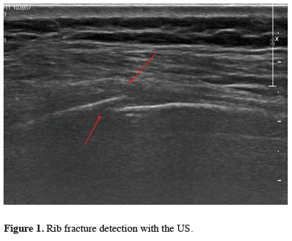
The chest x-ray (Samsung, GC80, Korea) was performed without a grid nearly 180 cm in posterioanterior position with a 85 kVp and 3.2 mAs. Ultrasonography was performed on patients by the radiologist using a 5-12 MHz linear array probe. Ultrasonography was performed longitudinally in such a way that the patient was in a lying or sitting position, starting from the place where the physical examination was most painful, including the adjacent ribs. During the ultrasound examination, cortical border irregularity, acoustic edge shadow, and local hematoma criteria were taken into consideration (Figure 1). Ultrasonography and chest radiography results were compared.
)
 Click Here to Zoom |
Figure 1: Rib fracture detection with the US. |
Statistical Analysis
Data were analyzed retrospectively. The variables were reported as mean and standard deviation (SD) with a confidence interval (CI) of 95%. Mean values were compared with the Student’s T-test and one-way analysis of variance (ANOVA). The ratios of categorical variables were given in percentages and compared with Fisher’s Exact test or Pearson’s chi-square test. In addition, sensitivity and specificity values are given as a result. The significance level was accepted as p < 0.05. All analyses were performed using SPSS 22.0 (SPSS Inc. Chicago, Illinois, USA) software.
Results
)
Table 1: Characteristics of the study group.
While the rib fractures were detected in 24 patients with chest x-ray, US detected rib fractures in 47 patients. The total number of fractures detected by chest x-ray was 37, and the total number of fractures detected by the US was 94. As a result of the comparison between the two examinations, the superiority of the US in the diagnosis of rib fracture was statistically significant compared to the chest radiography (p < 0,001) (Table 2).
)
Table 2: Detection of the rib fracture.
In the rib fracture diagnosis, the sensitivity of chest x-ray and US were 48.9% and 97.9%; specificity was 90.9% and 90.9%; positive predictive value (PPV) was 95.8% and 97.9%; negative predictive value (NPV) 29.4% and 90.9%; the accuracy rate was 56.9% and 96.6%, respectively (Table 3).
)
Discussion
Ultrasound examination has become popular in recent years as a useful and non-invasive diagnostic tool [5]. There are studies in which ultrasonography has better results than chest x-ray in the diagnosis of rib fractures [4,6,10,11]. In a meta-analysis, the specificity and sensitivity of the US were reported to be better [12]. Studies are reporting that ultrasound is also useful in sternal fractures [13,14]. Chan reported that the US has higher accuracy in thoracic bone fractures [7]. The sensitivity, specificity, and efficacy values of ultrasound in the diagnosis of rib fracture were found to be better than chest radiography in our study in accordance with the literature. Our patients were admitted to the outpatient clinic an average of 4.3 days after trauma. Hwang et al reported that no significant effect on the results that duration time until the US examination [5].
Being non-invasive, portable, relatively inexpensive, non-radiation and reproducible are the most critical advantages of ultrasonography and these make ultrasonography a valuable diagnostic tool [1]. It is also a reliable diagnostic tool for pregnant patients [8]. A plain chest x-ray may overlook uncalcified rib fractures, especially in costal cartilages [3]. It is known that ultrasound is more sensitive than plain x-ray and tomography in such fractures [8]. Studies are showing that ultrasound could better detect costal cartilage fractures and costochondral junction fractures [4,10]. Griffith et al [10] reported that plain chest radiography could detect rib fractures in 12% of patients with chest trauma, while ultrasonography could detect in 78% and ultrasonography was more sensitive. In our study, rib fracture was detected in 41.4% of patients with chest x-ray and 81% with the US. Martino et al [15] argued that ultrasonography is more useful in rib and costal cartilage fractures and should be performed in every patient with chest trauma as it could detect internal organ injuries.
Accurate identification of rib fractures by ultrasonography in case of clinical suspicion usually provides some advantages in practice, although it does not substantially alter patient management. First, with the early diagnosis of rib fractures, it might be necessary to initiate an appropriate pain management protocol that confirms the use of analgesic therapy or intercostal nerve blockages. Medical advice on employee return to work and activities is essential [7]. Meanwhile, the US is an easy, accessible, rapid, and accurate diagnostic procedure in hemodynamically unstable patients with hemothorax and/or pneumothorax [1]. The diagnosis of rib fracture is also important in terms of the medico-legal perspective [2,16]. The significant ultrasonography disadvantages are the difficulty in diagnosis in large-breasted patients, obesity, fractures in the ribs under the scapula, or the first rib fractures below the clavicle [1,5,8].
In conclusion, we believe that preferring ultrasound instead of thorax CT for its less radiation and lower cost at the next step beyond chest x-ray for patients, who applied to the outpatient clinic with minor trauma and whose general condition is well, is more beneficial. With more studies including more centers and the number of patients, ultrasonography would be used more widely as a result of supporting the effectiveness of ultrasonography in the diagnosis of rib fracture.
Declaration of conflicting interests
The authors declared no conflicts of interest with respect to the authorship and/or publication of this article.
Funding
The authors received no financial support for the research and/or authorship of this article.