
Abstract
Introduction
Case Presentation
)
 Click Here to Zoom |
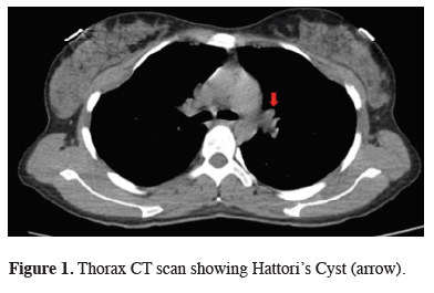
Figure 1: Thorax CT scan showing Hattori’s Cyst (arrow). |
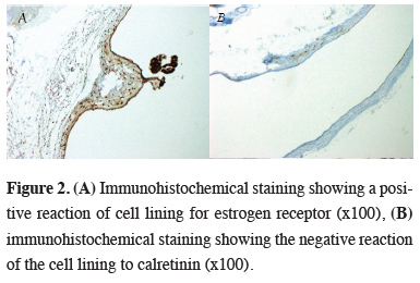
Surgical resection was planned. The patient underwent video-assisted thoracoscopic surgery (VATS). On exploration, a lesion of 5x5 cm, filled with serous fluid, on the left paracardiac region was seen. The cyst showed invasion to the mediastinal pleura; however, it had no invasion to the lungs. Total excision of the cyst was performed. Postoperative histopathological examination showed the cyst wall consisted of smooth muscle and connective tissues lined by ciliated epithelium with no cytological atypia. Immunohistochemical examination showed a positive reaction for Estrogen Receptor (ER) and Wilms’ Tumor Gene 1 (WT-1), and negative for Calretinin (Figures 2a, b). Based on these findings, the patient was diagnosed with Hattori’s Cyst of the mediastinum.
)
 Click Here to Zoom |
Figure 2: (A) Immunohistochemical staining showing a positive reaction of cell lining for estrogen receptor (x100), (B) immunohistochemical staining showing the negative reaction of the cell lining to calretinin (x100). |
Written informed consent was obtained from the patient for publication of her data.
Discussion
Regarding mediastinal cysts, a decision to operate is frequently made if the patient is symptomatic. As the size of the cyst increases, symptoms (chest pain, shortness of breath, palpitations, coughing, etc.) due to compression increase as well. Follow-up in the outpatient clinics is usually recommended to those patients with asymptomatic, small cysts [4]. Since our patient had symptoms of chest pain, and the cyst was 5 cm, we decided to perform surgery.
To date, 26 cases of Mediastinal Hattori’s Cyst have been published in the literature, including our case. In these cases, the size, location, and symptoms caused by the cyst were investigated, and it was determined that cysts were more common in the left thoracic cavity compared to the right [5].
The cyst of Mullerian origin in the mediastinum is still unclear. The reason for the cysts being at the thoracic location is because of the developmental misplacement of the endosalpingeal or endo-salpingeal epithelium. One theory is that coelomic epithelium develops on the cranial end of the plica mesonephrica at the level of the third to fifth thoracic vertebral blastema [6]. All of the cases published in the literature define the cyst in the posterior mediastinum and paravertebral area. The case we present is the first case of a cyst of embryonic Mullerian origin in the anterior mediastinum.
The prognosis of all cases of Hattori’s Cyst published to date has been shown to be good. Recurrence after surgical resection has not been reported. We performed total excision of the cyst in order to prevent a recurrence. It is difficult to make a preoperative diagnosis; therefore, it is essential that the differential diagnosis of these cysts should be made after resection. This way differentiation of these cystic tumors from bronchogenic or neurogenic cysts can be achieved. Detailed histopathological and immunohistochemical evaluation is imperative for definitive diagnosis.
Previously published 25 cases refer to Hattori’s Cyst in the posterior mediastinum. Our case shows that Hattori’s Cyst can also be seen in the anterior mediastinum and should be included in the differential diagnosis. Further clinical studies are needed to have more information on this subject.
Declaration of conflicting interests
The authors declared no conflicts of interest with respect to the authorship and/or publication of this article.
Funding
The authors received no financial support.
Authors’ contributions
MSOM; Collected the data, performed the analysis, co-wrote the paper, CBS; performed the analysis, co-wrote the paper, VE, MM; co-wrote the paper, NU; contributed data/analysis tools.