
Abstract
Introduction
Accompanied by the literature, we aimed to present a destroyed lung case caused by CLL’s mass effect in the posterior mediastinum, since it is infrequent.
Case Presentation
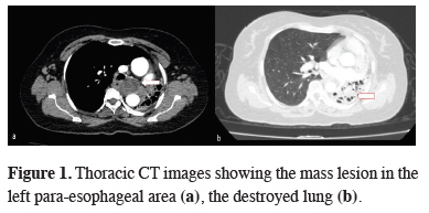
The second application of the patient to our clinic was 5 years later with complaints of shortness of breath, cough and chest pain. In her anamnesis, she had a history of tuberculosis during the childhood period. At the physical examination, respiratory sounds could not be detected on the left hemithorax and there was no palpable lymphadenopathy at any other part of the body. The sedimentation rate was 25mm/h. The other laboratory parameters were in normal ranges except mild leukocytosis. There was no growth in the tuberculosis culture of the bronchial sample taken from the patient. Abdominal ultrasonography was normal. Computed tomography of the thorax revealed a well-circumscribed, 46x33 mm mass lesion in the left para-esophageal area, springing the descending aorta to the left and compressing the left main bronchus. The left lung was destroyed, mediastinal shift was apparent together with the hyperaeration of the right lung (Figure 1).
)
 Click Here to Zoom |
Figure 1: Thoracic CT images showing the mass lesion in the left para-esophageal area (a), the destroyed lung (b). |
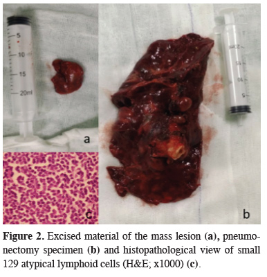
During the fiberoptic bronchoscopy, a long segment external compression was detected starting from the trachea’s distal level descending to the left main bronchus. Diffuse edema and abundant mucopurulent secretion was present in the left main bronchus. Pseudomonas aeroginosa was reproduced in the culture. Antibiotic treatment of piperacilin-tazobactam in compliance with the culture taken was initiated. Respiratory function test results were as follows; FEV1: 1.07 (51%), FVCex: 1.57 (57%), FEV1/FVC: 76% (96%). According to lung ventilation-perfusion scintigraphy results, no functional lung tissue was reported in the left lung area and the patient was appropriate for left a pneumonectomy. With the present findings, an operation was planned for both the diagnosis and treatment. Mediastinal mass excision and pneumonectomy was performed via left thoracotomy. The patient, was discharged on the 5th postoperative day without any complications. The histopathological diagnosis was bronchiectasis, and the mediastinal mass was compatible with chronic lymphocytic leukemia (Figure 2). The patient was referred to the oncology clinic for further treatment.
)
 Click Here to Zoom |
Figure 2: Excised material of the mass lesion (a), pneumonectomy specimen (b) and histopathological view of small 129 atypical lymphoid cells (H&E; x1000) (c). |
Written informed consent was obtained from the patient for publication of her data.
Discussion
Differential diagnosis is important in patients with mediastinal mass. For this reason, establishment of the diagnosis by preoperative PET/CT and a tissue biopsy is recommended [5]. Such as, a diagnosis of lymphoma obtained by biopsy will direct the primary treatment of the patients mostly medically. In our case, the mediastinal mass was suitable for a transbronchial biopsy and PET-CT could have been done. However, the patient with the destroyed lung had a definitive indication for a surgical intervention so the former interventions had not been performed. Left pneumonectomy and mediastinal mass excision were performed for definite diagnosis and treatment.
CLL patients may be asymptomatic at the time of diagnosis or present with B symptoms (weight loss, fever, night sweats), pallor caused by anemia, hemorrhage due to thrombocytopenia, symptoms due to lymph node or organ involvement, and frequent infections because of hypogammaglobulinemia. The most common finding on physical examination is lymphadenopathy (LAP), and splenomegaly. Hepatomegaly can also be seen [6]. Thoracic complications are not uncommon in hospitalized patients with CLL, but the specific etiology and incidence of these complications are limited [7]. Our case had nonspecific symptoms such as cough and chest pain at the time of admission. When the literature is reviewed, CLL cases with bronchial compression due to mass effect after mediastinal involvement and causing destroyed lung are rare [8].
In conclusion, the coexistence of CLL, which causes bronchiectasis and destroyed lung by compressing the bronchi with mass effect, is rare. Hematological malignancies should be kept in mind in the presence of bronchiectasis and mediastinal mass for which surgery is considered.
Declaration of conflicting interests
The authors declared no conflicts of interest with respect to the authorship and/or publication of this article.
Funding
The authors received no financial support.
Authors’ contributions
CH, KCC; conceived and designed the current case report. FC; collected the clinical data, AGY; examined the pathological preparates. KCC, CH, FC; co-wrote the paper. All authors discussed the case with under the literature data and contributed to the final manuscript.