
Abstract
Introduction
)
Table 1.Types of hypoplasia and characteristics in pulmonary hypoplasia [4].
This case presents a patient diagnosed with bilateral pulmonary lower lobe hypoplasia during surgery, who subsequently underwent bilateral lower lobectomy.
Case Presentation
)
 Click Here to Zoom |
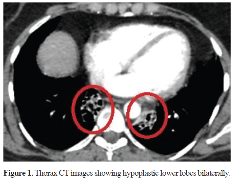
Figure 1: Thorax CT images showing hypoplastic lower lobes bilaterally. |
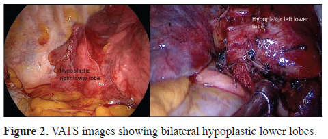
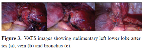
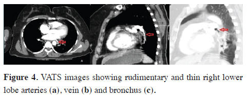
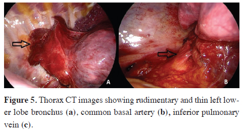
During exploration (first left, then right lower lobectomy via uniportal VATS, with one month between the two surgeries), both bilateral lower lobes were found to be completely rudimentary (Figure 2). The left lower lobe vein and bronchus were hypoplastic. The left lower lobe common basal segment artery was thin, and the superior segment artery was not visible (Figure 3). The right lower lobe vein was also hypoplastic, and the common basal and superior pulmonary artery branches were not visible. The right lower lobe bronchus was extremely narrow and rudimentary, similar to the left lower lobe bronchus (Figure 4).
)
)
)
 Click Here to Zoom |
Figure 2: VATS images showing bilateral hypoplastic lower lobes. |
 Click Here to Zoom |
Figure 3: VATS images showing rudimentary left lower lobe arteries (a), vein (b) and bronchus (c). |
 Click Here to Zoom |
Figure 4: VATS images showing rudimentary and thin right lower lobe arteries (a), vein (b) and bronchus (c). |
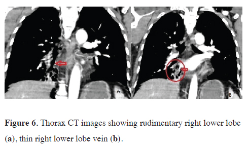
Postoperatively, detailed evaluation of pulmonary CT angiography revealed rudimentary continuation of the lower lobe branches of the venous, arterial, and bronchial systems toward the rudimentary lower lobes (Figures 5,6). The patient was discharged at day 3 uneventfully. Written informed consent was obtained from the patient for the publication of her data.
)
)
 Click Here to Zoom |
Figure 5: Thorax CT images showing rudimentary and thin left lower lobe bronchus (a), common basal artery (b), inferior pulmonary vein (c). |
 Click Here to Zoom |
Figure 6: Thorax CT images showing rudimentary right lower lobe (a), thin right lower lobe vein (b). |
Discussion
Congenital anomalies affecting other systems, such as cardiovascular, gastrointestinal, and genitourinary anomalies, may accompany pulmonary hypoplasia, leading to childhood diagnoses. In the absence of accompanying anomalies, patients may remain asymptomatic [7,8]. Diagnosis is usually incidental on chest radiographs, with the left side more commonly affected in adults. Complications like pneumothorax, pulmonary arterial hypertension, hemoptysis, or recurrent infections with bronchiectasis affecting the hypoplastic lung may necessitate hospital admission [9-11].
Radiological imaging typically reveals volume loss in the affected lobe or lung, mediastinal shift towards the hypoplastic side, volume increase in the contralateral lung or adjacent lobes, and elevation of the adjacent diaphragm [11,12]. CT scans are particularly effective for evaluating the diameter of tracheobronchial and major vascular structures, as well as extensive lung tissue damage, including damage to alveolar and bronchial structures. Hypoplastic bronchi can also be visualized on bronchoscopy [3].
Treatment in adults often involves conservative approaches such as managing infections, symptomatic relief with bronchodilators or expectorants, and prophylactic vaccination if necessary. However, in some patients, recurrent infections and cystic degeneration of the airways may necessitate surgical intervention, particularly if complications such as hemoptysis develop [5]. Minimally invasive surgical methods are preferred, with thoracotomy reserved for cases where thoracoscopic exposure is not feasible due to excessive adhesions [13]. In this patient, recurrent pulmonary infections and recent onset hemoptysis were the primary indications for surgical treatment, and bilateral thoracoscopic lower lobectomy was performed.
In conclusion, pulmonary hypoplasia is rare in adults and typically presents with recurrent lower respiratory tract infections and associated complications. Anatomical surgical resection is recommended using minimally invasive methods and/or thoracotomy
Declaration of conflicting interests
The authors declared no conflicts of interest with respect
to the authorship and/or publication of this article.
Funding
The authors received no financial support.
Authors’ contributions
MGP,VI,YB: conceived and designed the current case
report, co-wrote the paper, collected the clinical data.
The authors discussed the case under the literature data
together and constituted the final manuscript.
Reference
This article is an open access article distributed under the terms and conditions of the Creative Commons Attribution (CC BY) license (http://creativecommons.org/licenses/ by/4.0/).