
Abstract
Materials and Methods: Data from 7 patients who underwent VATS lung-sparing airway resections in our clinic between February 2022 and December 2024 were retrospectively evaluated in terms of surgical indications, as well as preoperative, intraoperative, and postoperative conditions.
Results: The surgical procedures included isolated carinal resection and reconstruction in two patients, main bronchial bronchotomy with bronchoplastic resection and reconstruction in two patients, segmental sleeve resection of the right main bronchus in one patient, and segmental sleeve resection of the bronchus intermedius in two patients, including one posterior biportal and one anterior uniportal approach. The mean age was 46.4 ± 17.1 years (range: 26-67). Pathological diagnoses included four typical carcinoid tumors, one glomus tumor, one sarcoma, and one squamous cell carcinoma. All resections achieved negative surgical margins. Postoperatively, the mean time to chest drain removal was 1.86 ± 0.38 days, and the mean length of hospital stay was 4.43 ± 1.40 days. No intraoperative mortality occurred, and no major postoperative complications or anastomotic failures were observed.
Conclusions: VATS lung-sparing tracheobronchial and carinal resections are feasible and safe in carefully selected patients. These procedures allow complete oncological resection while preserving lung parenchyma, with favorable perioperative outcomes and short hospital stays. Minimally invasive lung-sparing airway surgery may offers a valuable alternative to more extensive resections in selected tracheobronchial tumors.
Introduction
Historically, the standard approach to access the carina, right main bronchus, and bronchus intermedius has been via a right posterolateral thoracotomy through the fourth or fifth intercostal space [5]. Recently, with growing experience in VATS, these complex procedures have become feasible in experienced centers [2]. VATS has demonstrated clear advantages over open surgery, including reduced postoperative pain, shorter hospital stays, decreased morbidity, and improved cosmetic outcomes [6].
This study presents technical details of seven cases: two isolated carinal resections with reconstruction due to malignancies localized to the carina; one isolated right main bronchus resection and sleeve anastomosis for a glomus tumor in the right main bronchus; two bronchoplastic resections and reconstructions of the main bronchus for typical carcinoid tumors, one in the right main bronchus and the other in the left main bronchus; and two segmental sleeve resections of the bronchus intermedius for typical carcinoid tumors confined to that region.
Materials and Methods
Preoperative evaluation
A comprehensive preoperative evaluation was performed
to assess surgical eligibility and to plan the operative
strategy. Contrast-enhanced thoracic computed
tomography (CT) was used to define tumor location,
size, anatomical relationships, and possible invasion
of adjacent structures. Positron emission tomographycomputed
tomography (PET-CT) was performed to
evaluate metabolic activity and to identify regional
lymph node involvement and distant metastases. Rigid
or fiberoptic bronchoscopy (FOB) was utilized for direct
visualization of the lesion, histopathological confirmation
via biopsy, and airway patency restoration when
required. Pulmonary function was assessed using forced
expiratory volume in one second (FEV1), with diffusing
capacity for carbon monoxide (DLCO) measured in
selected patients. Only patients deemed suitable from
both respiratory and cardiological perspectives were
considered candidates for surgical intervention.
Perioperative evaluation andsurgical technique
All patients were positioned in lateral decubitus. Selective
intubation was utilized in cases requiring main
bronchus or bronchus intermedius resection, while in
patients undergoing isolated carinal resection, singlelumen
intubation in combination with either an endobronchial
blocker or high-frequency jet ventilation
(HFJV) was employed. In one segmental sleeve resection
of the bronchus intermedius, a uniportal technique
was used, whereas a biportal approach was adopted in
all other cases. A 30º thoracoscope was used for visualization,
and a soft tissue retractor such as Alexis® (Applied
Medical Resources Corporation, Rancho Santa
Margarita, CA, USA) was placed in the utility incision
to facilitate retraction. Following intubation, all patients
underwent fiberoptic bronchoscopy (FOB) to reassess
the tumor localization and the planned surgical intervention.
In long-segment resections of the tracheobronchial
tree, release maneuvers are recommended to reduce
anastomotic tension [7]. In a total of 5 cases involving
isolated carinal sleeve resections, segmental sleeve resections
of the intermediate bronchus, and main bronchus
sleeve resections, the inferior pulmonary ligament
was released as a releasing maneuver. In the first case of
isolated carinal resection and reconstruction, additional
distal tracheal mobilization was performed. To further
reduce tension at the anastomotic site and prevent involuntary neck extension, the chin was sutured to the
manubrium of the sternum in both cases of isolated carinal
resection/reconstruction. This technique reduced
anastomotic tension and mitigated the risk of sudden
neck hyperextension. These sutures were removed after
approximately one week. To achieve optimal exposure
and facilitate anastomosis in procedures involving the
carina and right main bronchus, the azygos vein was
divided, and both ends were anchored to the chest wall
and mediastinal pleura.
In bronchoplastic resections, the tumor-containing airway segment was removed via bronchotomy, and the bronchial defect was repaired with primary sutures. In sleeve resections, anastomosis was started at the membranocartilaginous junction of the proximal airway using an inside-to-outside suturing technique along the cartilaginous wall. To avoid suture entanglement during thoracoscopic anastomosis, the first proximal stitch was passed through the parietal pleura, brought out through the utility incision, and temporarily secured under tension. After completion of two-thirds of the cartilaginous portion, the suture was retrieved, and the membranous portion was completed in the opposite direction. The anastomosis was finalized using an endoscopic knot pusher. For isolated carinal resections, the trachea was first partially anastomosed end-to-end with the left main bronchus. The right main bronchus was subsequently anastomosed endto- side to the remaining tracheobronchial opening.
Intraoperative frozen section analysis was performed in all patients to confirm negative bronchial margins. As no positive margins were identified on frozen section examination in any case, additional bronchial resection prior to anastomosis or bronchoplasty was not required. Additionally, to adhere to oncological principles and ease the anastomotic procedure, the right lower paratracheal and subcarinal lymph nodes were excised at this stage. To enhance anastomotic healing and prevent minor leaks, viable tissue flaps such as omentum, parietal pleura, or pericardial fat may be used. The application of fibrin sealants (e.g., Tisseel) over the anastomotic site is another option to prevent minor air leaks and reduce the risk of dehiscence. In our series, a parietal pleural flap was used in one case, and fibrin sealant (Tisseel) was applied in four cases.
Postoperative evaluation
Patients were extubated and monitored in the intensive care unit ICU for one day before transfer to the thoracic
surgery ward. Chest drains were removed when daily
drainage was <100 mL, no air leak was present, and
complete lung expansion was confirmed on posteroanterior
chest radiographs. Carinal resection patients were
monitored until chin sutures were removed. Outpatient
follow-up was scheduled 10 days post-discharge. No
FOB was required postoperatively in any case.
Results
The cohort consisted of seven patients with a mean age of 46.4 ± 17.1 years (range: 26-67). Pathological diagnoses included four typical carcinoid tumors, one glomus tumor, one sarcoma, and one squamous cell carcinoma. All resections achieved negative surgical margins.
Postoperatively, the mean time to chest drain removal was 2.00 ± 0.58 days, and the mean length of hospital stay was 4.43 ± 1.81 days. No intraoperative mortality occurred. No major or minor complications or anastomosis failures occurred in the postoperative period. The mean follow-up duration was 22 months, during which no local or distant recurrence was observed, and all patients were alive at the last follow-up.
Case 1. Right VATS bronchotomy and bronchoplastic
resection and reconstruction of the right main bronchus
A patient evaluated due to a typical carcinoid tumor
localized in the proximal right main bronchus underwent
FOB, which revealed a 1×1 cm lesion on the posterior
wall of the distal right main bronchus extending
toward the carina. The lesion was located in the right
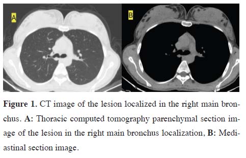
main bronchus on thorax CT (Figure 1). Selective intubation
was performed, and with the patient in the left
lateral decubitus position, thoracic access was achieved
through a 3 cm utility incision at the 4th intercostal space
in the anterior-mid axillary line. The camera port was
inserted through the 7th intercostal space along the posterior
axillary line. The surgical assistant retracted the
lung inferiorly using a lung retractor inserted through
the camera port to expose the paratracheal area. For exposure, the azygos vein was divided, and its posterior
stump was sutured to the chest wall. The hilar (10R)
lymph node was excised. The right main bronchus was
circumferentially dissected and suspended with a vessel
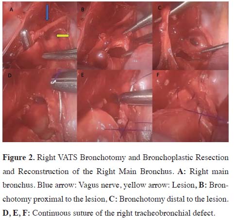
loop. Macroscopically, a bulging lesion over the membranous
portion at the carinal level was observed. The
distal trachea was mobilized close to the carinal plane,
with careful preservation of the vagus nerve. Using a
scalpel and endoscopic scissors, a bronchotomy was
performed from the distal right main bronchus toward
the carina, ensuring negative surgical margins, and the
lesion was excised over a 1 cm² area. The resulting defect
was closed with a continuous suture using 3-0 polypropylene
with a round needle. The anastomotic line
was reinforced with a parietal pleural flap, and fibrin
sealant (Tisseel) was applied to support the repair site
(Figure 2). The operation lasted 125 minutes with an
estimated blood loss of 100 mL. The chest drain was
removed on postoperative day 2, and the patient was
discharged on postoperative day 3.
)
)
 Click Here to Zoom |
Figure 1. CT image of the lesion localized in the right main bronchus. A: Thoracic computed tomography parenchymal section image of the lesion in the right main bronchus localization, B: Mediastinal section image. |
 Click Here to Zoom |
Figure 2. Right VATS Bronchotomy and Bronchoplastic Resection and Reconstruction of the Right Main Bronchus. A: Right main bronchus. Blue arrow: Vagus nerve, yellow arrow: Lesion, B: Bronchotomy proximal to the lesion, C: Bronchotomy distal to the lesion. D, E, F: Continuous suture of the right tracheobronchial defect. |
i>Case 2. Left VATS left main bronchus bronchotomy
and bronchoplastic resection and reconstruction
Due to a 1 cm typical carcinoid tumor located in the
distal left main bronchus, surgery was performed in
the right lateral decubitus position under selective intubation.
Thoracic access was achieved through a 3 cm
utility incision at the 4th intercostal space in the midaxillary
line. The camera port was inserted at the 7th
intercostal space along the posterior axillary line. The
lung was retracted anteriorly to expose and dissect the
left main bronchus, which was circumferentially mobilized
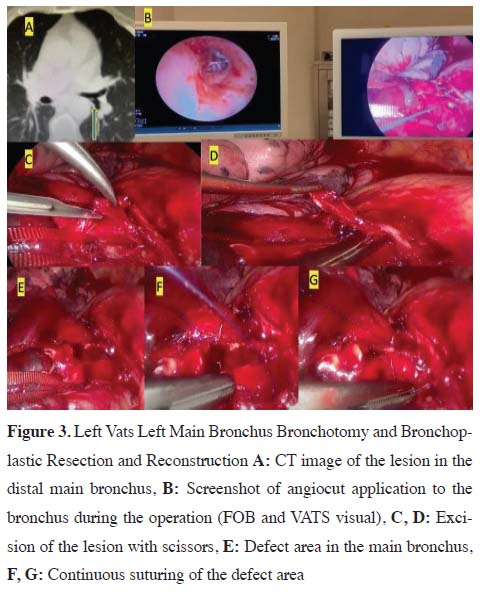
and suspended using a vessel loop. To localize
the lesion intraoperatively, an angiocatheter was inserted
intraluminally through the distal left main bronchus. The
catheter was visualized via FOB, confirming the lesion’s
position. A bronchotomy was performed on the membranous
portion of the left main bronchus using a scalpel, and
the lesion was resected with negative surgical margins.
The defect was closed with a continuous suture using 3-0
polypropylene with a double round needle. Lymph nodes
from the aortopulmonary window and subcarinal region
were excised. No air leak was observed during the leak
test. A single 28 Fr chest drain was placed through the
camera port, and the procedure was concluded. The operation
lasted 90 minutes with an estimated blood loss of
150 mL. The chest drain was removed on postoperative
day 3, and the patient was discharged on day 4 (Figure 3).
)
 Click Here to Zoom |
Figure 3. Left Vats Left Main Bronchus Bronchotomy and Bronchoplastic Resection and Reconstruction A: CT image of the lesion in the distal main bronchus, B: Screenshot of angiocut application to the bronchus during the operation (FOB and VATS visual), C, D: Excision of the lesion with scissors, E: Defect area in the main bronchus, F, G: Continuous suturing of the defect area |
Case 3. Right VATS segmental sleeve resection of the
right main bronchus
Due to a glomus tumor localized on the lateral wall of
the distal right main bronchus, surgery was performed
under selective intubation with the patient in the left lateral
decubitus position. Thoracic access was achieved
via a 3 cm utility incision at the 4th intercostal space.
A camera port was inserted at the 7th intercostal space
along the posterior axillary line. The azygos vein was
divided using an endoscopic stapler, and its ends were
anchored to the chest wall and mediastinal pleura to
provide adequate exposure. With careful preservation
of the vagus nerve, the trachea and right main bronchus
were dissected circumferentially and suspended using
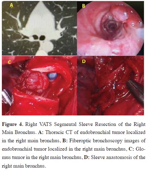
a vessel loop. A bronchotomy was performed with
a scalpel on the right main bronchus, allowing direct
visualization of the tumor. The affected segment of the
right main bronchus, including the lesion, was partially
resected, ensuring negative microscopic margins at
both proximal and distal ends. An end-to-end anastomosis
was then performed using a continuous suture
technique with 3-0 polypropylene and a double round
needle. After confirming the absence of an air leak, the
divided ends of the azygos vein were reapproximated
and sutured over the anastomotic line to provide additional
support. A single 28 Fr chest drain was placed in
the thoracic cavity. The operation lasted 130 minutes
with an estimated blood loss of 250 mL. The chest drain
was removed on postoperative day 2, and the patient
was discharged on day 3 (Figure 4).
)
 Click Here to Zoom |
Figure 4. Right VATS Segmental Sleeve Resection of the Right Main Bronchus. A: Thoracic CT of endobronchial tumor localized in the right main bronchus, B: Fiberoptic bronchoscopy images of endobronchial tumor localized in the right main bronchus, C: Glomus tumor in the right main bronchus, D: Sleeve anastomosis of the right main bronchus. |
Case 4. Right VATS isolated carina resection and reconstruction
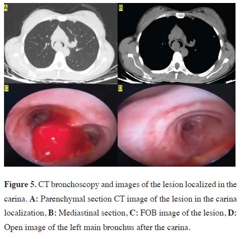
Rigid bronchoscopy was performed by the interventional
pulmonology team due to a follicular dendritic cell
sarcoma localized at the carina. The lesion was causing
near-total obstruction of the right main bronchus and
approximately 70% obliteration of the left main bronchus
(Figure 5). Airway patency was partially restored
via argon plasma coagulation (APC), and definitive
surgical resection was planned and performed 10 days
later. Under general anesthesia, the patient was placed
in the left lateral decubitus position. Endotracheal intubation
was performed with an 8.0-mm tube, and an
endobronchial blocker was positioned in the right main
bronchus. A 3 cm utility incision was made through the
4th intercostal space, and the camera port was placed at
the 7th intercostal space along the posterior axillary line.
The azygos vein was divided using an endovascular
stapler to facilitate exposure. The trachea was circumferentially
mobilized and suspended with a vessel loop.
The main pulmonary artery and the superior pulmonary
vein were dissected, looped, and retracted inferiorly to
expose the right main bronchus and carina. The right
main bronchus was then dissected and suspended with a
vessel loop. Macroscopically, the tumor was protruding
approximately 2 cm outward from the membranous portion
at the posterior aspect of the carinal level. The right
main bronchus was first transected at its most proximal
margin using a scalpel. The left main bronchus was then
visualized and dissected distally, circumferentially mobilized,
and suspended with a vessel loop. For traction,
a 2-0 polyglactin was placed on the cartilaginous wall
of the left main bronchus. The distal trachea was transected
at the level corresponding to the proximal carina
using a scalpel. Before this step, the patient’s oxygen
saturation was optimized, and the endobronchial blocker
was withdrawn. Finally, using the traction suture
placed on the left main bronchus, the left main bronchus
was transected with a scalpel, and the specimen
was removed en bloc. At this stage, the patient's ventilation
was maintained via a high-frequency jet ventilation
(HFJV) cannula inserted from the trachea into the left
main bronchus. The inferior pulmonary ligament was
released, and the head was placed in flexion. End-toend
anastomosis between the left main bronchus and the
trachea was initiated using 3- 0 polypropylene with a
double round needle in a continuous suture technique.
The suture was advanced over approximately 1 cm, after which both ends were temporarily secured by tying
them to newly placed anchoring sutures, intentionally
leaving a gap at the medial aspect of the anastomosis.
Subsequently, a parabolic (crescent-shaped) cartilage
segment was excised from the tracheal edge at the remaining
gap to create an adequate opening for the endto-
side anastomosis of the right main bronchus. The
right main bronchus was then anastomosed to this site
using continuous sutures with 3-0 polypropylene and a
double round needle. No air leak was observed. For reinforcement,
fibrin sealant (Tisseel) was applied to the
anastomotic site. A single 28 Fr chest drain was placed
in the thoracic cavity. To reduce tension on the anastomosis
postoperatively, the patient’s chin was sutured to
the sternum using a No. 1 silk suture. The total operative
time was 4 hours, with an estimated blood loss of
350 mL. The chest drain was removed on postoperative
day 2. On day 7, the chin suture was removed, and the
patient was discharged.
)
 Click Here to Zoom |
Figure 5. CT bronchoscopy and images of the lesion localized in the carina. A: Parenchymal section CT image of the lesion in the carina localization, B: Mediastinal section, C: FOB image of the lesion, D: Open image of the left main bronchus after the carina. |
Case 5. Right VATS isolated carina resection and reconstruction
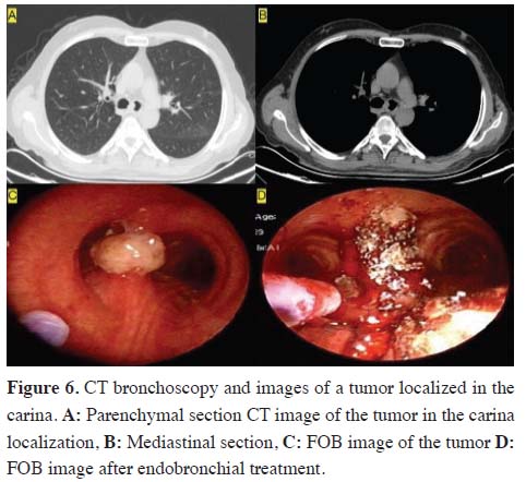
A patient diagnosed with squamous cell carcinoma localized
at the carina underwent surgery in the left lateral
decubitus position. The lesion was seen at the carina
level in the thorax CT and FOB examination (Figure
6). The patient was intubated with an 8.5 mm endotracheal
tube. Using FOB through the tube, an endobronchial
blocker was placed into the right main bronchus. A
working incision was made at the 4th intercostal space
along the mid-axillary line. The camera port was inserted
through the 8th intercostal space at the posterior axillary line. Through the camera port, the surgical assistant
used a lung retractor to displace the lung inferiorly,
exposing the paratracheal region. The azygos vein was
dissected, suspended, and divided using an endovascular
stapler. The distal trachea was circumferentially
mobilized and suspended with a vessel loop. Subsequently,
the right main bronchus was also dissected and
suspended. With the aid of FOB, the proximal resection
margin on the right main bronchus was identified. At
this point, the endobronchial blocker was withdrawn,
and bilateral ventilation was achieved using low tidal
volumes. The right main bronchus was transected with
negative surgical margins. The left main bronchus was
dissected distally, and a 2-0 polyglactin traction suture
was placed to facilitate exposure and manipulation. The
left main bronchus was transected near the carina with
negative surgical margins. Ventilation was maintained
via cross-field ventilation, using a sterile 5.5 mm spiral
endotracheal tube introduced directly into the left main
bronchus through the operative field. The carina was
then resected from the distal trachea using a scalpel, ensuring
negative surgical margins. The excised specimen
was sent for frozen section analysis, which confirmed
margin negativity. To reduce tension at the anastomotic
site, the inferior pulmonary ligament was released, and
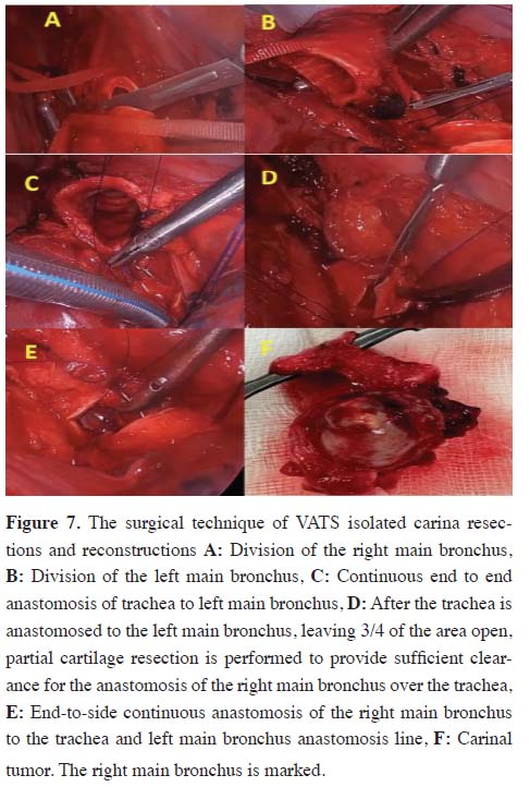
the patient’s head was placed in neck flexion. Reconstruction
began with an end-to-end anastomosis of the
left main bronchus to the trachea. The first stitch was
placed using 3-0 polypropylene with a double round needle,
starting from the junction between the cartilaginous
and membranous portions of the trachea (inside-out),
and continuing at the corresponding site on the left main
bronchus (outside-in), near the junction of the cartilage
and membranous wall. The end-to-end anastomosis was
completed, leaving a 1 cm gap on the medial aspect.
The free suture ends were tied to newly placed sutures
and then cut. To create a suitable opening for the right
main bronchus, a crescent-shaped piece of cartilage was
excised from the tracheal side. The right main bronchus
was then anastomosed end-to-side to this newly formed
opening. Near completion of the anastomosis, a caliber
mismatch was noted, prompting the excision of an additional
small cartilage segment from the tracheal edge to
ensure proper alignment. After confirming the absence
of air leaks, fibrin sealant (Tisseel) was applied to the
anastomotic site for reinforcement, and the ends of the
divided azygos vein were reapproximated and sutured
together. To prevent inadvertent neck hyperextension, the chin was sutured to the manubrium of the sternum
using No. 1 silk suture. A single 28 Fr chest drain was
placed. The total operative time was 180 minutes, and
the estimated blood loss was 350 mL. The chest drain
was removed on postoperative day 2, and the chin sutures
were taken down on day 7, at which point the patient
was discharged (Figure 7).
)
)
 Click Here to Zoom |
Figure 6. CT bronchoscopy and images of a tumor localized in the carina. A: Parenchymal section CT image of the tumor in the carina localization, B: Mediastinal section, C: FOB image of the tumor D: FOB image after endobronchial treatment. |
 Click Here to Zoom |
Figure 7. The surgical technique of VATS isolated carina resections and reconstructions A: Division of the right main bronchus, B: Division of the left main bronchus, C: Continuous end to end anastomosis of trachea to left main bronchus, D: After the trachea is anastomosed to the left main bronchus, leaving 3/4 of the area open, partial cartilage resection is performed to provide sufficient clearance for the anastomosis of the right main bronchus over the trachea, E: End-to-side continuous anastomosis of the right main bronchus to the trachea and left main bronchus anastomosis line, F: Carinal tumor. The right main bronchus is marked. |
Case 6. Right VATS segmental sleeve resection of the
bronchus intermedius: posterior approach
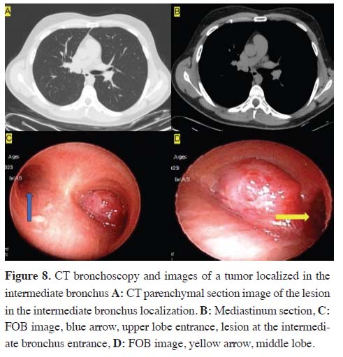
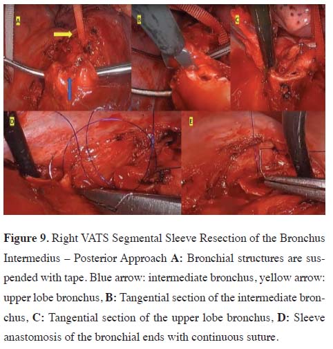
A patient diagnosed with a carcinoid tumor localized in
the right bronchus intermedius underwent surgery in the
left lateral decubitus position under selective intubation.
The lesion was seen in the intermediate bronchus in the
thorax CT and FOB examination (Figure 8). A 3 cm utility
incision was made through the 4th intercostal space along
the posterior axillary line. The camera port was placed at
the 7th intercostal space, also along the posterior axillary
line. The mediastinal pleura was incised posteriorly, and
dissection was performed down to the distal right main
bronchus and the right lower lobe bronchus to fully expose
the bronchial anatomy. Care was taken to avoid injury
to the pulmonary artery during dissection. The right
upper lobe bronchus and the middle lobe bronchus were
mobilized proximally, and the bronchus intermedius was
circumferentially dissected and suspended using a vessel
loop. Intraoperative FOB evaluation revealed that the
lesion extended from the posterior wall of the bronchus
intermedius at the level of the upper lobe bronchus distally.
Therefore, to preserve the upper lobe and ensure
negative margins, a curved (tangential) incision rather
than a linear one was made proximally on the bronchus
intermedius. The upper limit of the incision extended up
to the posterior wall of the upper lobe bronchus. A similar
curved incision was then made at the proximal portion of
the middle lobe bronchus, and the bronchus intermedius
was resected en bloc. Frozen section analysis confirmed
tumor-free surgical margins. To reduce tension on the
anastomosis, the inferior pulmonary ligament was released.
The bronchial reconstruction was performed with
a continuous end-to-end anastomosis using 3-0 polypropylene
with a double round needle (Figure 9). The procedure
lasted 130 minutes, and the estimated blood loss
was 200 mL. The chest drain was removed on postoperative
day 1, and the patient was discharged on day 3.
)
)
 Click Here to Zoom |
Figure 8. CT bronchoscopy and images of a tumor localized in the intermediate bronchus A: CT parenchymal section image of the lesion in the intermediate bronchus localization. B: Mediastinum section, C: FOB image, blue arrow, upper lobe entrance, lesion at the intermediate bronchus entrance, D: FOB image, yellow arrow, middle lobe. |
 Click Here to Zoom |
Figure 9. Right VATS Segmental Sleeve Resection of the Bronchus Intermedius – Posterior Approach A: Bronchial structures are suspended with tape. Blue arrow: intermediate bronchus, yellow arrow: upper lobe bronchus, B: Tangential section of the intermediate bronchus, C: Tangential section of the upper lobe bronchus, D: Sleeve anastomosis of the bronchial ends with continuous suture. |
Case 7. Right VATS segmental sleeve resection of the
bronchus intermedius: anterior uniportal approach
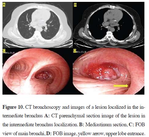
A patient diagnosed with a typical carcinoid tumor localized
in the bronchus intermedius underwent surgery in
the left lateral decubitus position under selective intubation.
The tumor was seen in the intermediate bronchus in
the thorax CT and FOB examination (Figure 10). Thoracic
access was achieved through a 3 cm uniportal utility
incision made at the 5th intercostal space along the
mid-axillary line. The procedure began with the opening
of an incomplete fissure to access the basal pulmonary
artery. The lung was then retracted anteriorly, and
the posterior mediastinal pleura was incised to expose
and excise a paraesophageal lymph node. Using a tunneling
technique posterior to the bronchus intermedius,
the dissection proceeded through the posterior mediastinal
pleura to reach the oblique fissure, which was divided
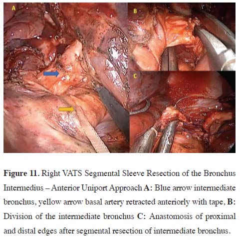
using an endoscopic stapler. Through the fissure,
the basal segmental pulmonary artery was looped with
a vessel tape and retracted anteriorly, allowing exposure
of the bronchus intermedius. The bronchus intermedius
was carefully dissected and encircled using a dissector.
It was transected just distal to the upper lobe take-off.
To ensure complete resection of the tumor, an additional
proximal ring of the bronchus intermedius was excised.
Macroscopically, the tumor showed protrusion through
the membranous wall toward the extrabronchial space;
this extension was dissected bluntly and sharply. After
proximal resection of the bronchus intermedius, subcarinal
and paraesophageal lymph nodes were removed. The distal bronchus intermedius, including the proximal portion
of the middle lobe bronchus, was also resected. Frozen
section analysis confirmed negative margins at both
proximal and distal ends. Bronchial reconstruction was
performed with a continuous anastomosis using 3-0 polypropylene
with a double round needle. During the leak
test, a small air leak was observed at the junction of the
cartilaginous and membranous portions on the posterior
side of the anastomosis. This defect was repaired with an
additional 3-0 polypropylene suture, and no further air
leak was detected upon retesting (Figure 11). The operation
lasted 180 minutes, with an estimated blood loss of
270 mL. The chest drain was removed on postoperative
day 2, and the patient was discharged on day 4.
)
)
 Click Here to Zoom |
Figure 10. CT bronchoscopy and images of a lesion localized in the intermediate bronchus A: CT parenchymal section image of the lesion in the intermediate bronchus localization. B: Mediastinum section, C: FOB view of main bronchi, D: FOB image, yellow arrow, upper lobe entrance. |
 Click Here to Zoom |
Figure 11. Right VATS Segmental Sleeve Resection of the Bronchus Intermedius – Anterior Uniport Approach A: Blue arrow intermediate bronchus, yellow arrow basal artery retracted anteriorly with tape, B: Division of the intermediate bronchus C: Anastomosis of proximal and distal edges after segmental resection of intermediate bronchus. |
Discussion
Declaration of conflicting interests
The authors declared no conflicts of interest with respect
to the authorship and/or publication of this article.
Funding
The authors received no financial support for the research
and/or authorship of this article.
Ethics approval
The study was approved by the Ethics Committee of
Dr. Ismail Fehmi Cumalıoglu City Hospital (2024/107).
Authors’ contribution
MEF: contributed to the definition of intellectual content,
VE: was involved in the manuscript preparation
and design, MÜ: contributed to the statistical analysis,
design, and editing, ACK: was involved in the clinical
studies, EK: contributed to the data acquisition, MSOM:
contributed to the editing, NY: contributed to the definition
of intellectual content, AÇ: was involved in the
literature search, MM: contributed to the review of the
manuscript, all authors have read and approved the final
version of the manuscript
Reference
This article is an open access article distributed under the terms and conditions of the Creative Commons Attribution (CC BY) license (http://creativecommons.org/ licenses/by/4.0/)