
Abstract
Materials and Methods: Patients who underwent surgery for lung cancer were retrospectively reviewed. Those with a postoperative histopathological diagnosis of LCNEC were included. Demographic and clinicopathological characteristics were analyzed, and factors affecting survival were investigated.
Results: Between January 2016 and December 2025, pulmonary resection was performed in 1066 patients with primary lung cancer, of whom 36 (3.4%) had LCNEC. The mean overall survival (OS) was 56.4 ± 8.0 months, with a 5-year OS rate of 42.8%. The mean disease-free survival (DFS) was 37.7 ± 7.4 months, with a 5-year DFS rate of 26.1%. Patients who received adjuvant therapy had significantly better OS (p < 0.001) and a trend toward improved DFS (p = 0.215) compared to those who did not, particularly in stage II and higher disease (p < 0.001 and p = 0.177, respectively). The presence of spread through air spaces (STAS) and higher N stage were associated with worse survival outcomes, although these differences did not reach statistical significance.
Conclusions: Adjuvant therapy is associated with improved survival in patients undergoing surgery for LCNEC.
Introduction
Under the World Health Organization (WHO) classification of lung tumors, LCNEC is categorized within the neuroendocrine neoplasms group, alongside typical carcinoid, atypical carcinoid, and SCLC. Compared to carcinoid tumors, LCNEC exhibits a higher Ki-67 proliferation index and increased mitotic counts. This profile is directly associated with tumor aggressiveness and a poor prognosis [2]. The Ki-67 index, even when obtained from small biopsies, serves as a crucial diagnostic aid for neuroendocrine tumors. Due to its aggressive nature and high propensity for early metastasis, LCNEC is classified as a high-grade neuroendocrine tumor, similar to SCLC [3]. In light of these characteristics, adjuvant therapy is recommended to prevent recurrence and improve overall survival [4].
Due to the rarity of pulmonary large cell neuroendocrine carcinoma (LCNEC), the number of studies focusing on this tumor is limited. In this study, we aimed to analyze the 10-year surgical outcomes of patients who underwent surgery for LCNEC, and to evaluate their demographic characteristics as well as tumor-related clinicopathological factors.
Materials and Methods
Patients who underwent surgery for lung cancer at our clinic were retrospectively reviewed. Among these cases, those whose postoperative pathology reports identified LCNEC or an LCNEC component were included in the study. Patients who did not undergo therapeutic surgery or those with incomplete medical records were excluded. This study was approved by the Ethics Committee of Kartal Dr. Lütfi Kırdar City Hospital (Approval No: 2026/010.99/26/1, date: 01.04.2026) and was conducted in accordance with the principles of the Declaration of Helsinki. Informed consent was obtained from each participant.
Data collection
The demographic characteristics of the included patients, the type of surgical treatment performed, tumor invasion parameters, PET/CT findings, the administration of adjuvant therapy, staging according to the 9th edition of the TNM system [5], overall survival, and disease-free survival were evaluated. Overall survival (OS) and disease-free survival (DFS) were defined as the time from the date of surgery to the date of death or recurrence, respectively.
Preoperative period
All patients who underwent surgical treatment for lung cancer at our institution were preoperatively evaluated using computed tomography (CT) and positron emission tomography (PET/CT). Patients presenting with lesions suggestive of primary lung malignancy on PET/ CT without evidence of mediastinal lymph node involvement or distant metastasis underwent direct surgical intervention. In these cases, wedge resection followed by intraoperative frozen section examination was performed and upon confirmation of malignancy, subsequent anatomical resection was carried out. For central lesions and tumors larger than 3 cm, transthoracic needle aspiration biopsy (TTNA) was utilized as the initial diagnostic modality. Invasive mediastinal staging, via endobronchial ultrasonography (EBUS) or mediastinoscopy, was indicated for tumors exceeding 3 cm in diameter. Patients with confirmed single-station N2 disease on preoperative invasive staging proceeded to surgery following neoadjuvant therapy, whereas those with multi-station N2 involvement were excluded from surgical resection [6]. Patients who received neoadjuvant therapy due to pN2 disease and were downstaged to cN0 in the preoperative period were included in the study. Functional status was evaluated through pulmonary function tests and echocardiography, while preoperative risk stratification was documented using the Charlson Comorbidity Index.
Surgical approach
The extent of anatomical resection comprising lobectomy, bilobectomy, sleeve lobectomy, or pneumonectomy was determined based on tumor size and localization. Complete macroscopic tumor clearance was achieved in all patients. In cases with chest wall involvement, additional en bloc chest wall resection was performed. Furthermore, systematic mediastinal lymph node dissection was carried out in every patient to ensure accurate pathological staging.
Postoperative follow-up
Patients who underwent surgical resection for LCNEC were monitored with thoracic CT scans every 3 months for the first 2 years and subsequently every 6 months for up to 5 years. While adjuvant therapy is conventionally recommended in routine NSCLC practice for patients with tumors larger than 4 cm or those with N1/N2 disease, in our study cohort, adjuvant treatment was recommended for LCNEC patients starting from Stage I. Adjuvant therapy was not administered in cases of patient refusal or due to an inability to tolerate chemotherapy-related toxicities. If follow-up assessments suggested recurrence, further evaluation via PET/CT was conducted and biopsy procedures were performed when clinically indicated.
Statistical Analysis
Categorical variables were presented as frequencies and percentages. Continuous variables were expressed as mean ± standard deviation and compared using the Student’s t-test. Overall survival (OS) and disease-free survival (DFS) were estimated using the Kaplan–Meier method, with statistical differences assessed by the logrank test. Survival analyses are carried out after excluding operative mortality. A p-value of less than 0.05 was considered statistically significant. All statistical analyses were carried out using SPSS version 26.0 (IBM Corporation, Armonk, NY, USA).
Results
Demographic characteristics, surgical approaches, invasion parameters, and TNM stages of the patients are presented in Table 1.
)
The mean age of the patients included in the study was 62.8 ± 8.7 years and the average tumor size was 4.56 ± 2.33 cm. On PET/CT examination, the mean maximum standardized uptake (SUVmax) value was calculated as 11.9 ± 7.3. In the resected tumors, the mean Ki-67 proliferation index was found to be 72.6 ± 14.7.
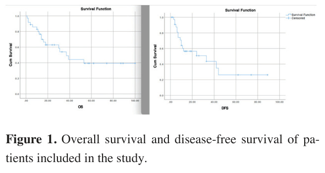
During the follow-up period, 19 patients (52.8%) remained alive, while 17 patients (47.2%) died; of these, 3 patients (8.3%) passed away within the first three postoperative months. The mean overall survival (OS) of the study cohort was 56.4 ± 8.0 months, with a 5-year OS rate of 42.8%. During the follow-up process, recurrence or metastasis was observed in 17 patients (47.2%). The mean disease-free survival (DFS) was 37.7 ± 7.4 months, and the 5-year DFS rate was found to be 26.1% (Figure 1). The sites of recurrence are presented in Table 2.
)
)
Analysis on spread through air spaces (STAS) reveals mean overall survival was 52.7 ± 9.6 months for STAS-positive patients, with a 5-year OS rate of 51.1%. For STAS-negative patients, mean OS was 61.5 ± 11.8 months, with a 5-year OS rate of 52.5% (p = 0.517). STAS-positive patients had a mean DFS of 40.4 ± 8.4 months and a 5-year DFS rate of 47.3%, whereas STASnegative patients exhibited a mean DFS of 39.0 ± 12.0 months, with a 5-year DFS rate of 22.2% (p = 0.910).
All patients included in the study were preoperatively staged as cN0. Occult hilar/mediastinal lymph node metastasis was observed in 11 patients (30.5%). In patients with mediastinal lymph node positivity, the mean overall survival was 36.4 ± 7.7 months, with a 5-year survival rate of 44.2%. For patients without mediastinal lymph node involvement, the mean overall survival was 56.7 ± 9.3 months, with a 5-year OS rate of 42.1% (p = 0.906). Regarding disease-free survival, the mean DFS for patients with mediastinal lymph node metastasis was 17.7 ± 2.8 months, with a 5-year DFS rate of 26.5%. In the group without mediastinal lymph node metastasis, these values were calculated as 40.2 ± 8.5 months and 29.5%, respectively (p = 0.769).
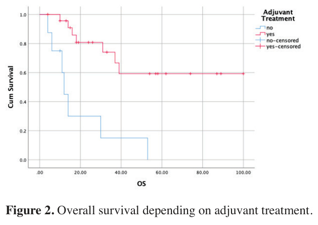
The impact of adjuvant therapy on mean survival values is detailed in Table 3 and illustrated in Figure 2.
)
)
 Click Here to Zoom |
Figure 1: Overall survival and disease-free survival of patients included in the study. |
 Click Here to Zoom |
Figure 2: Overall survival depending on adjuvant treatment. |
Table 1: Demographics of the patients, surgical approach, invasion parameters and staging.
Male
33 (91.66%) Female
3 (8.33%) Preoperative Biopsy
Malignant
24 (66.66%) LCNEC/NE
5 (13.88%) AdC
1 (2.77%) SqCC
2 (5.55%) Non-diagnostic
1 (2.77%) Not performed
3 (8.33%) Definitive diagnosis
Pure LCNEC
22 (61.11%) AdC component
8 (22.22%) SqCC component
4(11.11%) Pleomorphic C component
1 (2.77%) SCLC component
1 (2.77%) Symptoms
Incidental
9 (25.00%) Chronic cough
13 (36.11%) Fatigue
6 (16.66%) Hemoptisis
1 (2.77%) Dizziness
1 (2.77%) Surgery
RUL
11 (30.55%) RML
1 (2.77%) RLL
4 (11.11%) LUL
7 (19.44%) LLL
9 (25.00%) Lower bilobectomy
1 (2.77%) Right Pneumonectomy
1 (2.77%) Left Pneumonectomy
2 (5.55%) Incision
VATS
11 (30.55%) Thoracotomy
18 (50.00%) Conversion
7 (19.44%) Neoadjuvant Treatment
No
33 (91.66%) Yes
3 (8.33%) Mediastinal Staging
Not performed
13 (36.11%) EBUS
18(50.00%) Mediastinoscopy
5 (13.88%)
Negative
10 (27.77%) Positive
20 (55.55%) Not investigated
6 (16.66%) Pleural invasion
PL0
26 (72.22%) PL1
5 (13.88%) PL 2
0 (0.00%) PL 3
1(2.77%) Vascular invasion
No
21 (58.33%) Yes
15 (41.66%) Perineural invasion
No
32 (88.88%) Yes
4 (11.11%) T Status
T1b
5 (13.88%) T1c
7 (19.44%) T2a
8 (22.22%) T2b
4 (11.11%) T3
6 (16.66%) T4
6 (16.66%) N Status
N0
25 (69.44%) N1
7 (19.44%) N2a
3 (8.33%) N2b
1 (2.77%) M Status
M0
33 (91.66%) M1b
3 (8.33%) Stage
IA2
3 (8.33%) IA3
5 (13.88%) IB
5 (13.88%) 2A
3 (8.33%) 2B
9 (25.00%) 3A
5 (13.88%) 3B
3 (8.33%) IVA
3 (8.33%)
Abbrev.: AdC: Adenocarcinoma, EBUS: Endobronchial ultrasound, LCNEC/NE: Large cell neuroendocrine carcinoma/neuroendocrine carcinoma,
LLL: Left lower lobectomy, LUL: Left upper lobectomy, RLL: Right lower lobectomy, RML: Right middle lobectomy, RUL: Right upper lobectomy,
SCLC: Small Cell carcinoma, SqCC: Squamous cell carcinoma, STAS: Spread through air spaces, VATS: Video-assisted thoracoscopic surgery
Table 2: Recurrence sites of patients included in the study.
Recurrence Site
Lung
Mediastinum
Brain
Bone
Liver
Total
2 (5.55%)
7 (19.44%)
3 (8.33%)
4 (11.11%)
1 (2.77%)
17 (47.22%)
Table 3: Survival based on adjuvant treatment. DFS: Disease-free survival, OS: Overall survival.
Mean OS (months) based on presence of adjuvant treatment
Adjuvant (-)
Adjuvant (+)
p value
All stages
19.25±6.61
69.76±9.05
<0.0001
Stage 1
41.50±11.50
73.64±15.26
0.248
Stage 2-3-4
9.88±1.68
62.17±9.41
<0.0001
Discussion
Organization (WHO) classification [7]. With subsequent updates to immunohistochemical criteria, it was recategorized in 2015 within the neuroendocrine tumor group of the lung, along with typical carcinoid, atypical carcinoid, and small-cell lung cancer [8]. In the 2021 WHO classification, neuroendocrine neoplasms were categorized as low-grade (typical carcinoid), intermediate-grade (atypical carcinoid), and high-grade neuroendocrine carcinomas, the latter consisting of LCNEC and SCLC [9].
Despite improvements in the diagnostic yield of small-needle biopsies in recent years, establishing a preoperative diagnosis of LCNEC remains challenging using limited biopsy specimens. Due to the presence of excessive necrosis typically associated with these tumors, larger tissue samples are required to identify neuroendocrine morphology and perform specific immunohistochemical staining for neuroendocrine markers [10].
Consequently, patients with a final pathology of LCNEC are frequently preoperatively misdiagnosed as having
Mean DFS (months) based on presence of adjuvant treatment
Adjuvant (-)
Adjuvant (+)
p value
23.05±8.01
42.35±8.75
0.215
27.00±17.00
35.85±11.63
0.925
8.67±2.12
45.09±10.56
0.177 other types of NSCLC or SCLC [11,12]. In a study by Chen et al., it was reported that a diagnosis of LCNEC was achieved in only 6 out of 20 patients who underwent preoperative bronchoscopic or transthoracic biopsy [13]. Consistent with these findings, the majority of patients in our study could not be diagnosed with LCNEC preoperatively through small biopsies obtained via TTNA or transbronchial biopsy.
In patients with good performance status and adequate cardiopulmonary function, surgical intervention remains the primary therapeutic approach for earlystage LCNEC [14]. Previous studies have demonstrated that sublobar resections, such as segmentectomy and wedge resection, do not provide survival benefits comparable to those of lobar resections [15-18]. Due to the high growth potential of these tumors, extensive tumor necrosis and hemorrhage may be encountered intraoperatively [19], necessitating heightened caution, particularly when managing large masses. It has also been documented that spontaneous tumor rupture can lead to hemorrhagic shock [20]. At our clinic, the minimum extent of resection performed for all LCNEC patients was lobectomy. Our surgical experience indicates that these tumors contain significant necrosis and are prone to persistent, oozing-type bleeding due to excessive angiogenesis—though not necessarily massive hemorrhage. Consequently, this characteristic may contribute to an increased rate of conversion from video-assisted thoracoscopic surgery (VATS) to thoracotomy in this specific tumor type.
The optimal stage at which adjuvant therapy should be recommended for resectable LCNEC remains a subject of ongoing debate. Existing studies have reported benefits of postoperative adjuvant treatment in Stage I disease [21], Stage IB or higher disease, and Stage IIB or higher disease [15]. Although the number of studies on this topic is limited, the standard approach for early-stage LCNEC similar to that for SCLC is platinum-etoposide or irinotecan combinations. When all stages included in our study were analyzed, adjuvant therapy was found to provide a statistically significant benefit in OS. While a survival benefit was also observed in Stage I disease, it did not reach statistical significance. In contrast, for Stage II and higher disease, a statistically significant improvement in OS was observed. These findings suggest that while adjuvant therapy benefits the entire cohort, its efficacy is more pronounced in Stage II and higher disease. In DFS analyses, although adjuvant therapy appeared more beneficial, this advantage did not achieve statistical significance.
First described by Kadota et al. in 2015 [22], spread through air spaces (STAS) refers to clusters of tumor cells or single tumor cells within the air spaces beyond the edge of the main tumor. Although STAS was formally recognized as a prognostic feature only for nonmucinous adenocarcinoma in the 2021 WHO classification of lung tumors [23], it has been shown to adversely affect prognosis in other histopathological subtypes as well, despite its prevalence and prognostic significance being more extensively documented in adenocarcinomas [24-26]. In a study by Aly et al. focusing on STAS in neuroendocrine lung cancers, the prevalence of STAS in LCNEC was reported as 43%, and its presence was identified as a poor prognostic factor for both diseasefree survival and overall survival [27]. Consistently, our study suggested that STAS is a poor prognostic factor for LCNEC, although this association did not reach statistical significance.
Preoperative PET/CT is mandatory for patients with confirmed or suspected lung cancer. Invasive mediastinal staging is indicated when the tumor size exceeds 3 cm or if PET/CT reveals suspicious uptake in mediastinal or hilar lymph nodes [28]. Nodal upstaging is recognized as a poor prognostic factor in lung cancer. Previous studies have demonstrated nodal upstaging rates of 10% in clinical Stage I LCNEC and 8% in clinical Stage II LCNEC [15]. In our study, the rates of occult pN1 and pN2 disease were consistent with the literature. Furthermore, although the association did not reach statistical significance, nodal upstaging was found to be associated with a worse prognosis.
Ki-67 is an antigen expressed by the MKI67 gene and has been identified as a parameter associated with a poor prognosis [29]. In neuroendocrine lung tumors, the Ki-67 proliferation index is typically below 20% to 30% in typical and atypical carcinoids, whereas it often exceeds 50% and frequently surpasses 70% to 80% in LCNEC and SCLC [30]. Consequently, the Ki-67 index plays a pivotal role in refining the differential diagnosis of neuroendocrine neoplasms, particularly in small-needle biopsy specimens where tissue samples are limited.
Due to the rarity of LCNEC and the limited number of publications in the literature, reported survival rates vary within a wide range, despite the well-established aggressive nature and poor prognosis of this tumor. Studies have demonstrated that 5-year OS rates for all stages of LCNEC range from 18% [31] to 53.8% [32], and even in Stage I disease alone, these values fluctuate between 18% and 88%. These studies indicate that adjuvant therapy following surgery in resectable patients is associated with improved OS, with N and M stages being the most prominent factors influencing OS [13]. The number of publications reporting DFS rates for this tumor is considerably lower. One study reported a 5-year DFS of 32.2% and identified adjuvant chemotherapy and TNM stage as the primary factors affecting DFS [33].
In conclusion, our study reveals the administration of adjuvant therapy provides a statistically significant benefit in overall survival, it was also observed that STAS status and N status influence survival outcomes. These parameters were further found to be associated with disease-free survival.
Limitations of the study
Our study has several limitations. First is its retrospective design; however, clinical records at our institution were meticulously maintained and documented. The second limitation is the relatively small number of the sample size. This constraint is commonly encountered in the majority of existing literature on this specific subject. The primary reason why parameters such as STAS presence and N status did not reach statistical significance in their impact on overall and disease-free survival is likely the limited sample size. Nevertheless, this limitation is inherent to most studies focusing on this exceptionally rare tumor type.