
Abstract
Introduction
When risk factors for vascular migration are evaluated, increased intrathoracic pressure due to positivepressure ventilation, physical movements, sneezing, weightlifting, or coughing appears to be prominent. Vascular complications include arterial and venous injuries as well as hematoma formation. Vascular injuries are most frequently observed during femoral vein procedures and least commonly during subclavian vein access. It is thought that after guidewire placement, the dilator or catheter may damage the vessel wall, leading to bending of the guidewire and subsequent advancement toward the vessel wall [2].
In this case, no acute tracheobronchial injury was observed in the immediate post-catheterization period; rather, the complication developed because of the synergistic effect of chronic erosion of the bronchial wall by the distal tip of the port catheter and tissue necrosis caused by a superimposed invasive fungal infection (aspergilloma).
Tracheobronchial perforation is described in the literature as an early complication of catheterization, whereas delayed tracheobronchial perforation has been reported in only a few cases. To our knowledge, tracheobronchial perforation following port-related mediastinal aspergilloma has not been previously reported in the literature.
Case Presentation
)
 Click Here to Zoom |
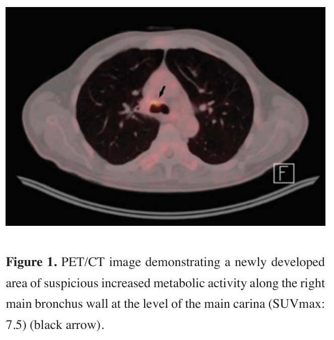
Figure 1: PET/CT image demonstrating a newly developed area of suspicious increased metabolic activity along the right main bronchus wall at the level of the main carina (SUVmax: 7.5) (black arrow). |
Due to suspicious imaging findings suggestive of an endobronchial lesion in a patient with malignancy, bronchoscopy was performed under general anesthesia. At the level of the main carina, a white, soft tissue lesion eroding the bronchial mucosa was identified at the origin of the right bronchial system, and biopsy specimens were obtained and sent for histopathological examination. In addition, a bronchoalveolar lavage sample from the right bronchial system was collected for further analysis. Fungal spores and hyphae were observed in both specimens, with Aspergillus species reported as the predominant organism. The biopsy specimens were also submitted for tissue culture, which yielded growth of Haemophilus parainfluenzae.
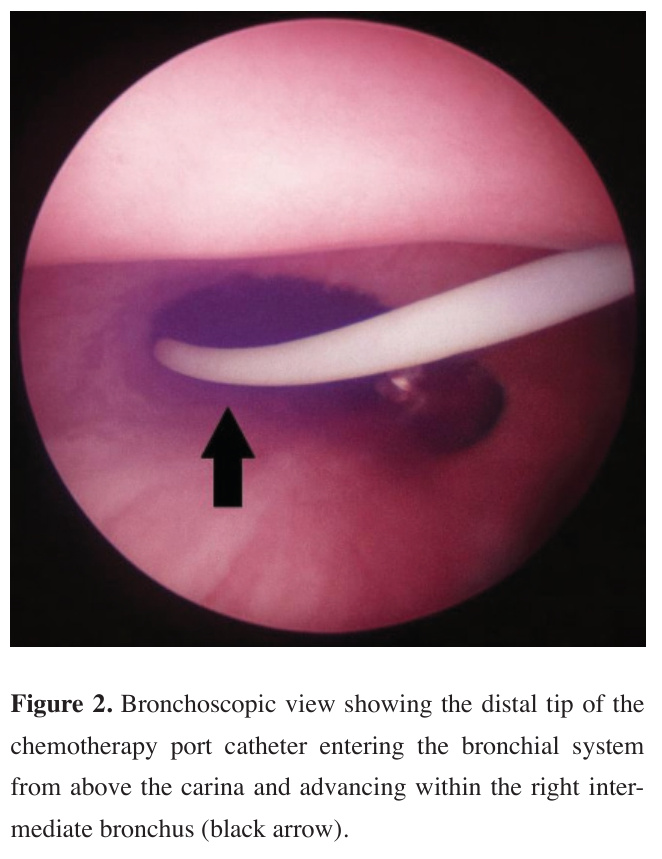
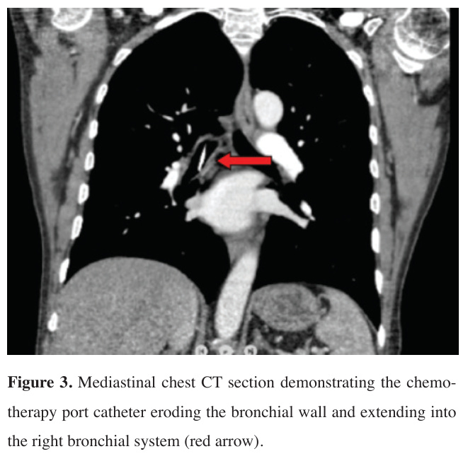
After seven weeks of antifungal therapy, the patient was re-evaluated with follow-up bronchoscopy under general anesthesia. During the procedure, the distal tip of the chemotherapy port catheter was observed to penetrate the mucosa and enter the right bronchial system (Figure 2). The site of penetration corresponded to the mucosal area where fungal growth had been identified during the previous bronchoscopy. The bronchoscopy was terminated, and a postoperative chest CT scan was subsequently performed (Figure 3). Imaging revealed the catheter component of the port within the right main bronchus. No pneumothorax or hemothorax was observed.
)
)
 Click Here to Zoom |
Figure 2: Bronchoscopic view showing the distal tip of the chemotherapy port catheter entering the bronchial system from above the carina and advancing within the right intermediate bronchus (black arrow). |
 Click Here to Zoom |
Figure 3: Mediastinal chest CT section demonstrating the chemotherapy port catheter eroding the bronchial wall and extending into the right bronchial system (red arrow). |
Interventional radiology and cardiovascular surgery departments were consulted, and surgical removal of the chemotherapy port under general anesthesia was decided. The patient was taken to the operating room with the involvement of these teams. During catheter removal, the patient was placed in the Trendelenburg position to prevent air embolism, and bronchoscopic access to the bronchial system was achieved through the endotracheal tube. To minimize the risk of endobronchial bleeding during catheter withdrawal, a bronchial blocker was introduced through the endotracheal tube and the balloon was inflated. While the right main bronchus was monitored bronchoscopically, the surgical field was prepared in a sterile manner, and the chemotherapy port was removed via surgical incisions.
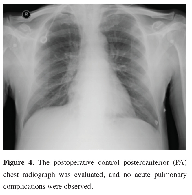
The patient was extubated and transferred to the ward with stable vital signs and no perioperative complications. Postoperative posteroanterior chest radiography revealed no complications (Figure 4). After five days of clinical, laboratory, and radiological follow-up, the patient was discharged. Written informed consent was obtained from the patient for the use of the medical data in academic studies.
)
 Click Here to Zoom |
Figure 4: The postoperative control posteroanterior (PA) chest radiograph was evaluated, and no acute pulmonary complications were observed. |
Discussion
The subclavian vein, internal jugular vein, and femoral vein can be used for central venous catheterization. The subclavian vein is frequently preferred for catheterization due to its lower risk of thrombosis, lower incidence of infection and relatively low rates of mechanical complications compared to other central venous access sites [4].
It has been observed that the rate of acute complications that may arise during ultrasound-guided central venous catheterization is lower compared to catheterization performed blindly [5]. Some of the possible complications include cardiac, mechanical, thrombotic, infectious, and pulmonary complications.
Among acute cardiac complications, arrhythmias are prominent, and this condition is most often associated with the guidewire contacting the right atrium. Fluoroscopy-assisted guidewire placement can help prevent vascular injury [6]. When vascular injury develops, most hematomas can be spontaneously resolved; however, in some cases, if progression or abscess formation occurs, evaluation should be performed with thoracic computed tomography, and drainage should be applied if hemothorax or empyema is detected [7].
Pulmonary complications include pneumothorax, pneumomediastinum, chylothorax, tracheobronchial injury, and air embolism [8]. Numerous catheter attempts and the use of an inappropriately sized catheter can increase the risk of pneumothorax. Tracheobronchial injuries can occur because of needle contact with the trachea or bronchus during vascular access attempts via guidewire [9]. Imaging methods that can aid in the diagnosis of pulmonary complications are plain radiography and CT scans.
In patients receiving mechanical ventilator support, small perforations in the endotracheal or tracheostomy cuff can lead to significant air leakage, requiring tube replacement. Especially in patients exposed to high airway pressure, holes under the cuff can cause pneumomediastinum or subcutaneous emphysema [10].
Air embolism can occur during catheter placement or irrigation. The negative intrathoracic pressure created during catheter placement can lead to air entry into the vessel. Massive air embolism can cause hypoxia. To prevent this, the patient should be placed in the Trendelenburg position, and all catheter access points should be closed. If air embolism is suspected, high-flow oxygen therapy should be administered [5,11].
In the management of these complications, simple catheter removal is theoretically considered an undesirable method as it can lead to venous air embolism or uncontrollable bleeding in the airway [12].
Early tracheobronchial perforation usually occurs within the first few days following an iatrogenic procedure and is mostly associated with technical errors, excessive mechanical trauma, or direct damage to the tracheal or bronchial walls during the procedure (e.g., intubation, bronchoscopy, central venous catheterization, etc.). Late tracheal perforation refers to cases where symptoms appear weeks or even months later; in these cases, processes such as tissue ischemia, chronic compression, or infection are more prominent than direct trauma [13].
Central and peripheral catheter insertions can cause tracheobronchial perforation through mechanisms such as indirect compression of the trachea or bronchus, or excessive compression of adjacent tissues. Incorrect positioning of catheter tips, especially with long-term use, increases the risk of chronic compression on the tracheobronchial wall and subsequent tissue erosion [14]. Major factors that increase the risk of developing latestage tracheobronchial perforation include long-term steroid use, existing infections, malnutrition, diabetes, and other immunosuppressive conditions.
Fungal tracheobronchitis is a fungal infection that invades the trachea and bronchi, more common in immunocompromised patients. These infections can directly target the tracheobronchial mucosa, leading to tissue necrosis. Although rare, the literature reports that ulcerative or pseudomembranous forms of fungal involvement are associated with subcutaneous emphysema and tracheobronchial perforation by disrupting the wall integrity [15].
The angiotropic property of Aspergillus species allows for direct tissue and vascular invasion. Particularly in immunosuppressed individuals, this leads to mucosal necrosis and erosion of the bronchial wall. It has been reported that fungal infection creates a "second hit" effect on top of the mechanical damage caused by the catheter itself [15].
In a study on port catheter migrations, migration was observed in 7 patients (incidence rate 2.3%) in a group of 298 patients [16]. Literature reviews show that tracheobronchial injuries can occur in the acute phase during central venous catheterization [17]. However, the damage observed in our case report did not develop in the acute phase during the central venous catheterization procedure but occurred due to erosion of the distal end of the port catheter in the wall and fungal infection in the chronic phase. Fungal organisms are rarely pathogenic in healthy individuals; however, they can become pathogenic in immunosuppressed groups, as in our patient. [18].
Our case of right main bronchus perforation that developed in the late period following central venous catheterization adds a new pathophysiological link to the known spectrum of complications of this procedure based on current literature. Especially its late onset suggests that the case may involve a more complex mechanism related to catheter placement, mechanical pressure, infectious processes, or individual patient factors. In this respect, the presented case points to a rare but clinically significant complication in the literature and provides a unique contribution to the literature by raising awareness about longterm follow-up of central venous catheterization.
In conclusion, central venous access procedures are among the frequently performed invasive procedures in hospitals. These procedures also carry complications, and detailed evaluation and information gathering are crucial for developing a management plan for these complications. Especially in patients with accompanying comorbidities, complex catheterization procedures may require a multidisciplinary approach. As presented in our case report, early detection of tracheobronchial perforation, a late complication of central venous catheterization, is as vital for the patient as its prevention. Since perforation occurring during port catheter use can lead to sudden respiratory symptoms, allergic reactions, and potentially fatal complications if left undetected, close clinical monitoring and careful evaluation of these patients are critical. While it is not possible to completely prevent complications, their prevention should be one of the clinician's primary goals.