
Abstract
Introduction
This report highlights the oncologic rationale for isolated sternal metastasectomy and addresses the critical technical considerations required when operating in the presence of indwelling breast prostheses.
Case Presentation
)
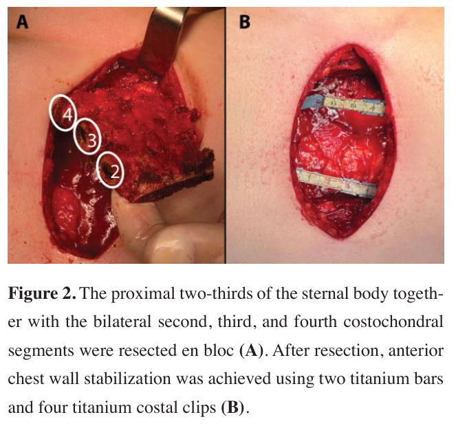
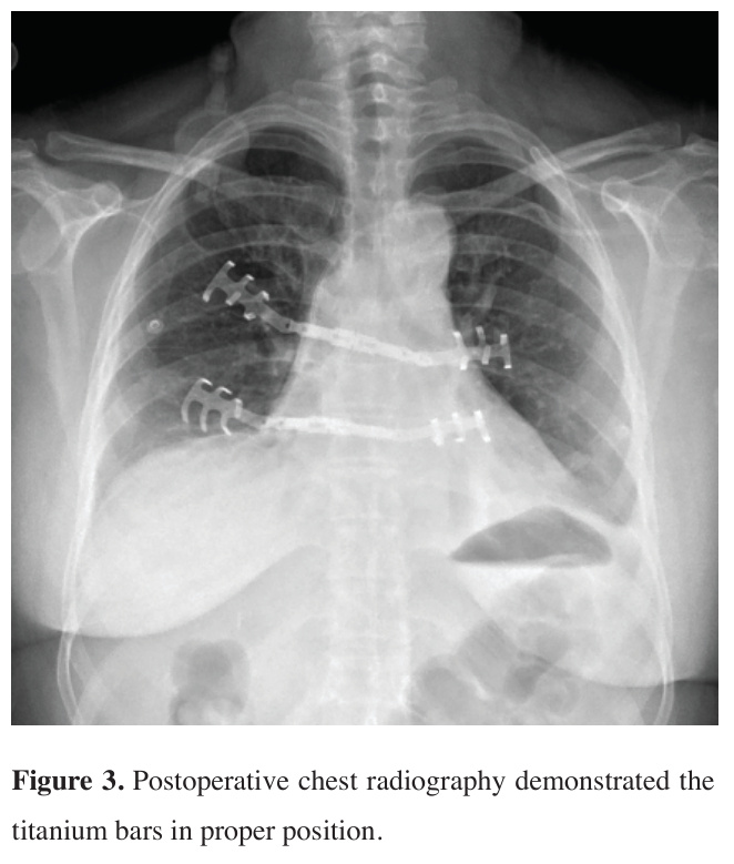
The patient was referred to our clinic by the oncology department for evaluation regarding metastasectomy. As the lesion was considered an isolated and resectable sternal metastasis, sternal resection and reconstruction were planned. Following a vertical midline incision of approximately 10 cm, the pectoralis major muscle was dissected, and the sternum along with the adjacent costochondral structures was mobilized. The proximal two-thirds of the sternal body together with the bilateral second, third, and fourth costochondral segments were resected en bloc. After resection, anterior chest wall stabilization was achieved using two titanium bars and four titanium costal clips (Figure 2). During placement of a clip at the left third costochondral region, injury to the existing breast implant was noted. Through the same surgical field, the deformed implant was removed, and after adequate irrigation and surgical field preparation, a new implant of the same size was placed in coordination with the plastic surgery team. The postoperative course was uneventful, and postoperative chest radiography demonstrated the titanium bars in proper position with no evidence of procedure-related complications (Figure 3). Accordingly, the patient was discharged on postoperative day three without any complications. Written informed consent for both participation and publication was obtained from the patient.
)
)
 Click Here to Zoom |
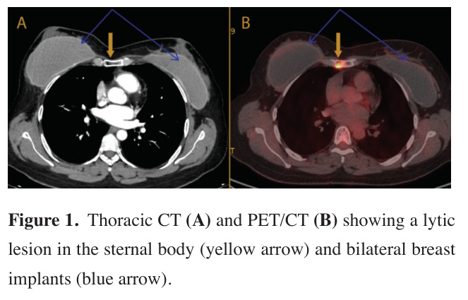
Figure 1: Thoracic CT (A) and PET/CT (B) showing a lytic lesion in the sternal body (yellow arrow) and bilateral breast implants (blue arrow). |
 Click Here to Zoom |
Figure 2: The proximal two-thirds of the sternal body together with the bilateral second, third, and fourth costochondral segments were resected en bloc (A). After resection, anterior chest wall stabilization was achieved using two titanium bars and four titanium costal clips (B). |
 Click Here to Zoom |
Figure 3: Postoperative chest radiography demonstrated the titanium bars in proper position. |
Discussion
Against this background, improved survival rates in breast cancer have made it necessary to offer breast reconstructive procedures to enhance quality of life and self-esteem. Implant-based breast reconstruction is the most utilized reconstructive approach following mastectomy; however, it is associated with a considerable risk of major complications and prosthesis failure, which may in turn complicate subsequent surgical interventions. Surgeons should be aware of possible implant injury and evaluate implant integrity intraoperatively [7]. In the study by Coombs et al. evaluating patients with preexisting breast implants who underwent cardiothoracic surgery or electrophysiologic procedures, postoperative implant-related complications within one year were reported in approximately 8%, 29%, and 5% of patients undergoing minimally invasive cardiac surgery (MICS), median sternotomy, and electrophysiologic procedures, respectively. Although the intraoperative implant rupture rate was higher in the MICS cohort, in 92% of cases the implant was safely manipulated through the original incision used for its placement [8]. These findings highlight the potential technical challenges posed by preexisting breast implants during thoracic procedures. In our case, the presence of bilateral breast implants posed a technical challenge during the mobilization of the costochondral structures and placement of clips through a limited incision. Despite prior bilateral implant-based reconstruction and adjuvant radiotherapy, the procedure was successfully completed with coordinated plastic surgery involvement. The implant damage was recognized intraoperatively at an early stage, and a new implant was inserted during the same session through the same operative field, without postoperative implant-related complications. Sternal resection and reconstruction performed without removal of the breast implant, using gentle and careful retraction and enlarging the surgical incision when necessary, may allow preservation of the integrity of the fibrous capsule and reduce the risk of postoperative incisional or prosthesis-related complications.
In conclusion, isolated sternal metastasis from breast cancer is an uncommon presentation in which surgical resection may be considered in carefully selected patients. However, prior implant-based breast reconstruction adds technical complexity to thoracic procedures. This case suggests that, with careful surgical planning and multidisciplinary collaboration, sternal metastasectomy and chest wall reconstruction can be safely performed despite intraoperative breast implant injury.