
Abstract
Introduction
The evolution from multiportal to uniportal VATS initially involved minor procedures; however, its indications subsequently expanded to include anatomical lung resections [2]. In parallel, robotic-assisted thoracic surgery (RATS) gained popularity because of its enhanced precision and ergonomics compared with VATS. Its advantages, including high-definition three-dimensional visualization and wristed instruments, facilitate meticulous dissection and effective lymphadenectomy [3].
Nevertheless, the conventional robotic approach typically requires multiple ports. To further minimize surgical trauma, many centers adopted the uniportal approach [4]. Several reports have demonstrated the technical feasibility of uniportal robotic-assisted thoracic surgery (U-RATS) using the da Vinci Xi platform. However, because the Xi system was not originally designed for uniportal access, technical challenges such as limited instrument triangulation and internal arm collisions may be encountered.
In contrast, the da Vinci SP system was specifically developed for single-port surgery. Its unique design allows triangulation of multiple instruments through a single access port, minimizes internal arm collisions, and incorporates a wristed camera that facilitates complex thoracic procedures [5].
Segmentectomy has been shown to provide oncological outcomes comparable to lobectomy in selected patients with early-stage lung cancer and also represents a valuable parenchyma-sparing option for benign lesions that are unsuitable for wedge resection [6,7]. Herein, we present a case of single-port robot-assisted common basal segmentectomy performed using the da Vinci SP platform for an indeterminate pulmonary nodule that was anatomically unsuitable for wedge resection.
Case Presentation
)
 Click Here to Zoom |
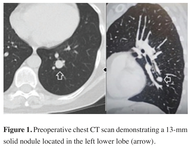
Figure 1: Preoperative chest CT scan demonstrating a 13-mm solid nodule located in the left lower lobe (arrow). |
Preoperative evaluation, including echocardiography and pulmonary function tests, revealed no significant abnormalities. Owing to the lesion’s central location and its proximity to the segmental bronchus, wedge resection was considered unsuitable. Therefore, a left lower lobe common basal segmentectomy was planned. Preparation of the single-port robotic system and docking
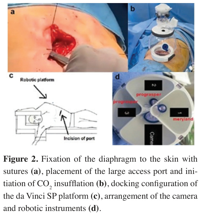
A 3.5-cm incision was made in the seventh left intercostal space. To optimize exposure, maintain diaphragmatic position, and facilitate instrument mobility, the diaphragm was sutured to the skin (Figure 2a). A large access port (2.7-7 cm) with a wound protector was inserted, and CO2 insufflation was initiated at a flow rate of 7 L/min (Figure 2b).
)
The robotic cart was positioned posterior to the patient, and docking was performed (Figure 2c). The procedure was initiated with the camera in the “below” position. A Maryland bipolar forceps and two Cadiere forceps were used as the working instruments (Figure 2d).
 Click Here to Zoom |
Figure 2: Fixation of the diaphragm to the skin with sutures (a), placement of the large access port and initiation of CO2 insufflation (b), docking configuration of the da Vinci SP platform (c), arrangement of the camera and robotic instruments (d). |
Surgical technique
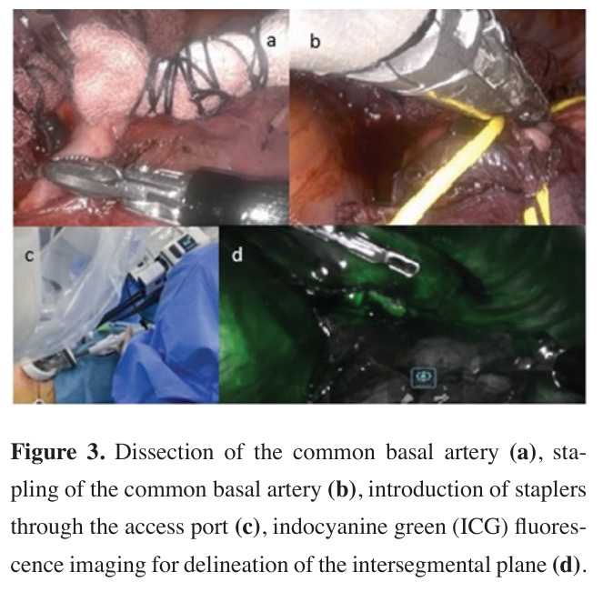
The operation commenced with division of the inferior pulmonary ligament and dissection of station 9 lymph nodes. Attention was then directed to the hilum, where the inferior pulmonary vein and subsequently the common basal vein were carefully dissected and exposed. The interlobar fissure was subsequently developed to identify and isolate the arterial branches supplying the basal segments. Station 11 lymph node dissection was performed, followed by completion of the anterior fissure. Then the common basal segment artery was stapled (Figures 3a,b).
)
During stapler application, one robotic arm was temporarily withdrawn to optimize instrument positioning and facilitate safe stapler deployment. The common basal vein and bronchus were divided using an endoscopic stapler. Although stapler insertion through the assistant port may be technically challenging, the use of a 12-mm ROSI port (Robotic operative suction and irrigation) provided adequate access, as the gray cap of the access port allows 270° rotation, thereby facilitating stapler positioning (Figure 3c).
Indocyanine green (ICG) fluorescence imaging was performed after intravenous administration of 0.5 mg/ kg ICG. The intersegmental plane between the preserved and devascularized lung parenchyma became clearly visible approximately 100 seconds after injection and was subsequently divided (Figure 3d).
 Click Here to Zoom |
Figure 3: Dissection of the common basal artery (a), stapling of the common basal artery (b), introduction of staplers through the access port (c), indocyanine green (ICG) fluorescence imaging for delineation of the intersegmental plane (d). |
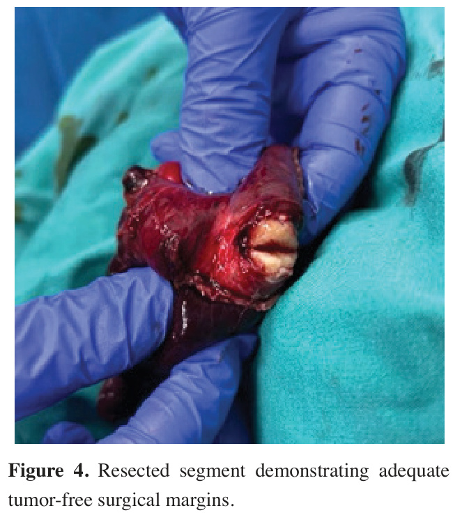
After specimen retrieval, meticulous hemostasis was achieved, intercostal nerve blocks were performed, and a chest tube was placed. Examination of the specimen confirmed that the lesion was located within the resected segment (Figure 4). Docking time was 15 minutes, and console time was 2 hours and 10 minutes. Estimated blood loss was 125 mL.
)
 Click Here to Zoom |
Figure 4: Resected segment demonstrating adequate tumor-free surgical margins. |
Postoperative course
The postoperative course was uneventful. The chest tube was removed on postoperative day 1, and the patient was discharged on postoperative day 3. Final pathological examination revealed a sclerosing pneumocytoma with a surgical margin of 3 mm. A total of six lymph nodes from stations 7, 10, and 11 were harvested, and no nodal involvement was identified. No postoperative complications occurred. At the 3-month follow-up, the patient remained well, with no evidence of recurrence or other complications.
Written informed consent was obtained from the patient for publication of this case report and accompanying images.
Discussion
Unlike the da Vinci Xi platform, which was adapted for uniportal procedures, the da Vinci SP system was specifically designed for single-port surgery. Its architecture allows triangulation of multiple instruments through a single access port while minimizing internal arm collisions. In addition, the wristed camera and flexible instrument deployment facilitate complex dissections and may expand the applicability of single-port robotic surgery to advanced thoracic procedures [5].
In the present case, a centrally located lesion involving the common basal segment of the left lower lobe precluded wedge resection. Therefore, anatomical segmentectomy was preferred to achieve complete resection while preserving functional lung parenchyma. Single-port robotic common basal segmentectomy enabled both definitive diagnosis and treatment in a single session, with minimal surgical trauma and an uneventful postoperative course.
Surgical resection remains the gold standard treatment for patients with early-stage and selected locally advanced non-small cell lung cancer (NSCLC), offering the best chance for cure when complete resection is achieved [9-12]. In this context, segmentectomy has been shown to provide oncological outcomes comparable to lobectomy in appropriately selected patients, without compromising survival or recurrence rates [13].
In recent years, an increasing number of small pulmonary nodules have been detected through lung cancer screening programs and widespread use of chest imaging during the COVID-19 pandemic [14,15]. Although many of these lesions are suspected to be malignant, a proportion are ultimately diagnosed as benign entities, including sclerosing pneumocytoma. In such cases, parenchyma-sparing anatomical resections may provide both definitive diagnosis and treatment while preserving pulmonary function. Furthermore, single-port robotic surgery is an effective option for these cases.