Current Thoracic Surgery
2026 , Vol 11 , Issue 2
Presternal elastofibroma: an unusual anterior chest wall presentation following median sternotomy
Current Thoracic Surgery 2026;11(2):199-202.
1Department of Thoracic Surgery, Faculty of Medicine, Gazi University, Ankara, Türkiye
DOI : 10.26663/cts.2026.028
SummaryIntroductionCase PresentationDiscussionReference
Summary
Elastofibroma is an uncommon benign soft-tissue lesion that most frequently arises in the subscapular region of elderly patients. It predominantly affects females, and occurrences outside the periscapular region are exceedingly rare and may pose a diagnostic challenge. A 59-year-old man presented with a progressively enlarging, palpable, and painful mass in the presternal region. Physical examination revealed a firm, fixed lesion beneath a previous sternotomy scar. Ultrasonography demonstrated a vascularized soft-tissue mass, while thoracic computed tomography showed a solid lesion adjacent to the sternum. Because malignancy could not be ruled out, complete surgical excision was performed. Histopathological evaluation established the diagnosis of elastofibroma. The postoperative course was uneventful, and no recurrence was detected during follow-up. Presternal elastofibroma is an exceptionally rare entity. In the present case, chronic mechanical irritation related to a malpositioned sternal wire may have contributed to lesion development, highlighting the importance of considering elastofibroma in the differential diagnosis of unusual anterior chest wall masses.
Introduction
Elastofibroma is a benign soft-tissue lesion that typically arises in the subscapular region in middle-aged to elderly individuals. It is characterized by slow growth, absence of a true capsule, and poorly defined margins. Although etiopathogenesis remains poorly understood, several mechanisms have been proposed. The most widely accepted hypothesis suggests that repetitive mechanical stress and microtrauma induce reactive hyperproliferation of elastic and collagen fibers [1]. Histopathologically, elastofibroma is an unencapsulated lesion composed of dense collagen bundles and thickened, fragmented elastic fibers arranged haphazardly among fibroblasts and adipocytes. While its borders may be partially discernible, they are typically irregular. Although the subscapular region is a classic location, atypical localizations have been reported rarely [2,3]. In this report, we present the clinical, radiological, and histopathological features of an elastofibroma located in the presternal region. We also discuss a potential etiopathogenetic mechanism involving chronic microtrauma from a sternal wire, in light of the existing literature.
Case Presentation
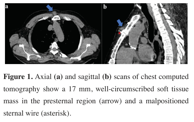
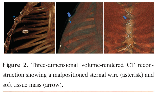
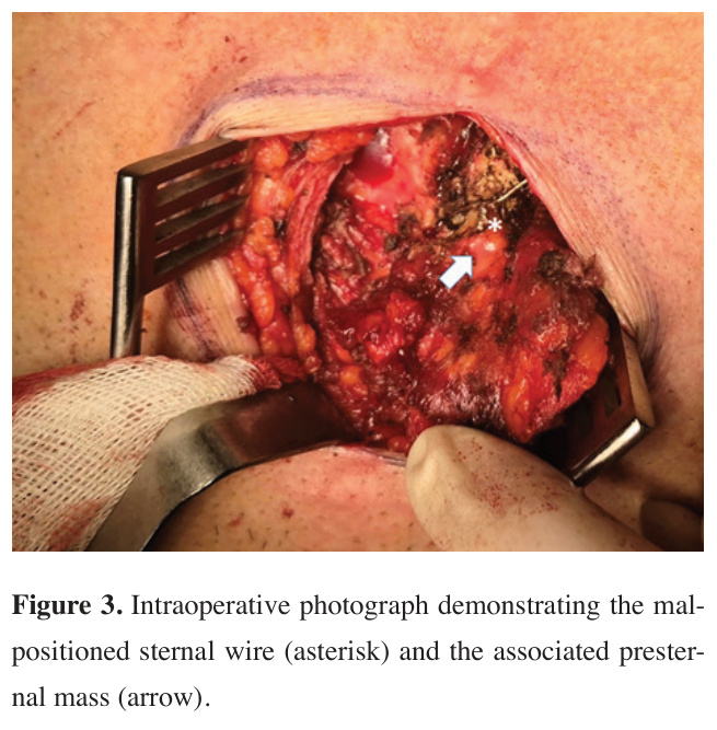
A 59-year-old man presented with a palpable mass in the presternal region that had been present for approximately one year and had progressively increased in size over recent months. His medical history included coronary artery bypass grafting and aortic–mitral valve replacement in 2005, left knee prosthesis surgery, hypertension, and coronary artery disease. On physical examination, a midline sternotomy scar was noted, along with a subcutaneous nodular lesion at the level of the manubriosternal junction measuring approximately 1.5-2 cm in diameter. The lesion was well-circumscribed, firm and elastic in consistency, fixed to the deep plane, and non-mobile. No erythema, local warmth, or ulceration of the overlying skin was observed. Ultrasonographic examination demonstrated a 15 × 14 mm nonspecific hypoechoic lesion with lobulated contours in the subcutaneous tissue, showing marked vascularity on Doppler imaging; therefore, a tissue diagnosis was recommended. Non-contrast thoracic computed tomography revealed a nodular softtissue density lesion measuring approximately 17 mm at the same level, accompanied by reticulonodular increased density in the surrounding subcutaneous tissue. No osseous erosion or invasion was identified (Figures 1,2). Given the atypical location and vascularity, malignancy could not be excluded, and surgical excision was planned. A 4-5 cm skin incision was made along the previous sternotomy scar. Following dissection through the skin and subcutaneous tissue, the mass was identified within the adipose tissue. A malpositioned sternal closure wire extending anteriorly into the lesion was observed. The lesion was dissected free and excised en bloc, and the twisted end of the sternal wire was bent back and repositioned (Figure 3). There were no intraoperative or perioperative complications. Gross examination revealed a 1.5 cm mass within a 4 × 5 cm specimen, with cystic areas containing fibrinoid material. Histopathological examination revealed an acellular, dense collagen matrix with thick, fragmented, eosinophilic elastic fibers interspersed throughout. Globules arranged linearly along these fibers were observed. Islands of mature lipocytes (fat deposits) were detected between the fibroelastic stroma. Immunohistochemical analysis demonstrated characteristic elastic fibers highlighted by Verhoeff-Van Gieson staining, confirming the diagnosis of elastofibroma. Postoperatively, the patient developed a wound seroma, which was managed with daily dressing changes without the need for surgical revision. Vital signs remained stable throughout the hospitalization. Anticoagulation therapy was carefully monitored, with warfarin dosage adjusted according to INR values and subcutaneous enoxaparin administered until a therapeutic INR level (>2.0) was achieved. The patient was subsequently discharged without any further complications. At 1-year follow-up, no recurrence was observed. Written informed consent for publication of the case report was obtained from the patient.


Click Here to Zoom
Figure 1: Axial (a) and sagittal (b) scans of chest computed tomography show a 17 mm, well-circumscribed soft tissue mass in the presternal region (arrow) and a malpositioned sternal wire (asterisk).


Click Here to Zoom
Figure 2: Three-dimensional volume-rendered CT reconstruction showing a malpositioned sternal wire (asterisk) and soft tissue mass (arrow).


Click Here to Zoom
Figure 3: Intraoperative photograph demonstrating the malpositioned sternal wire (asterisk) and the associated presternal mass (arrow).
Discussion
This case highlights an unusual presternal localization of elastofibroma, presenting as an anterior chest wall mass adjacent to a sternotomy site. Elastofibroma dorsi typically occurs in individuals over 50 years of age, most commonly between the sixth and seventh decades, and shows a female predominance. It is classically located between the inferior angle of the scapula and the thoracic wall. Occurrence in male patients and outside the periscapular region is considerably less common [3]. In this regard, our case is atypical in both patient demographics and lesion location. The etiology of elastofibroma is thought to involve repetitive mechanical stress and microtrauma, possibly in combination with genetic predisposition. Recent studies have also demonstrated cytogenetic abnormalities and clonal fibroblastic proliferation, suggesting that elastofibroma represents a lesion with intermediate features between reactive and neoplastic processes [1]. In the present case, the lesion developed in close association with a malpositioned sternal wire. We hypothesize that chronic microtrauma induced by the wire, which was exacerbated by respiratory motion and cardiac pulsation, may have triggered localized fibroelastic proliferation, analogous to the friction-related mechanism described in classical periscapular elastofibroma.

Radiological evaluation of elastofibroma typically involves ultrasonography, CT, and MRI. Classic imaging findings include a heterogeneous soft-tissue mass with attenuation similar to skeletal muscle and interspersed fatty streaks, producing a layered or striated appearance [4,5]. However, in atypical locations, these characteristic features may be absent. In our case, imaging findings were non-specific, and malignancy could not be excluded, necessitating surgical excision. In the differential diagnosis of the lesion, a vascularized soft-tissue malignancy was considered the most likely possibility. The painful nature of the lesion, its fixation to the surrounding tissues, and the marked vascularity observed on Doppler ultrasonography initially raised suspicion for a soft-tissue malignancy. Computed tomography findings further supported the presence of a soft-tissue mass. Granulation tissue associated with a malpositioned sternal wire was also included in the differential diagnosis. Other considerations included benign soft-tissue neoplasms, such as desmoid tumors and fibromas. As a definitive radiological diagnosis could not be established, histopathological examination was performed for definitive diagnosis.

Histopathological examination remains the gold standard for diagnosis, with characteristic findings including dense collagen bundles, fragmented elastic fibers, and interspersed adipose tissue. Special stains such as Verhoeff-Van Gieson are essential for confirming the diagnosis and excluding malignancy [6]. Although conservative management may be appropriate for asymptomatic lesions in typical locations, surgical excision is recommended in symptomatic cases or when malignancy cannot be ruled out. Recurrence after complete excision is exceedingly rare, consistent with the favorable outcome observed in our patient.

In conclusion, although elastofibroma is most commonly encountered in the periscapular region, fixed presternal lesions are usually regarded as potentially malignant. Therefore, complete surgical excision is often preferred to establish a definitive diagnosis and provide treatment. In our case, histopathological evaluation of a lesion that had been excised under the presumptive diagnosis of malignancy revealed an elastofibroma. Elastofibroma should be considered in the differential diagnosis of anterior chest wall masses in patients with a history of sternotomy, particularly when sternal closure wires are present. Awareness of this rare presentation may help avoid misdiagnosis and guide appropriate management.

Reference

1)

Nagano S, Yokouchi M, Setoyama T, Sasaki H, Shimada H, Kawamura I et al. Elastofibroma dorsi: Surgical indications and complications of a rare soft tissue tumor. Mol Clin Oncol 2014; 2: 421-4.

2)

Chandrasekar CR, Grimer RJ, Carter SR, Tillman RM, Abudu A, Davies AM et al. Elastofibroma dorsi: an uncommon benign pseudotumour. Sarcoma 2008; 2008: 756565.

3)

El Hammoumi M, Qtaibi A, Arsalane A, El Oueriachi F, Kabiri el H. Elastofibroma dorsi: clinicopathological analysis of 76 cases. Korean J Thorac Cardiovasc Surg 2014; 47: 111-6.

4)

Karakurt O, Kaplan T, Gunal N, Gulbahar G, Kocer B, Han S et al. Elastofibroma dorsi management and outcomes: review of 16 cases. Interact Cardiovasc Thorac Surg 2014; 18: 197-201.

5)

Tripathy S, Parida GK, Naswa N, Subudhi K, Sreedharan Thankarajan AR, Reddy S. Elastofibroma Dorsi: Findings on 18F-Fluorodeoxyglucose Positron Emission Tomography-Computed Tomography. Indian J Nucl Med 2019; 34: 258-9.

6)

Tasli F, Vardar E, Argon A, Kabat T, Deniz S, Nart A et al. Histochemical and immunohistochemical characteristics of elastofibromas. Pol J Pathol 2014; 65: 120-4.