
Abstract
Introduction
The severity of this condition varies; it is classified into cervical, cervicothoracic, thoracoabdominal, and abdominal subtypes, each requiring distinct management strategies. Ectopia cordis is frequently associated with additional anomalies, notably Cantrell’s pentalogy, which includes diaphragmatic hernia, omphalocele, sternal and pericardial defects, and congenital heart disease. The absence of pericardial protection further increases vulnerability to trauma and infection [2]. When the heart protrudes through the chest wall above the diaphragm, it is classified as extrathoracic ectopia cordis, one of the most severe forms of the anomaly. Prognosis is generally poor, particularly in thoracic ectopia cordis, emphasizing the need for immediate intervention [3].
Surgical reconstruction primarily involves repositioning the heart and covering the chest wall defect; however, because of the high mortality associated with this condition, successful surgical correction remains uncommon [4]. Single-stage thoracoabdominal repairs have demonstrated feasibility, reducing the need for multiple interventions. Given the complexity of ectopia cordis, surgical approaches must be tailored to the severity of the presentation and associated anomalies to optimize survival [3].
Although successful repairs have been reported, longterm outcomes and quality of life remain underexplored due to the rarity of this condition [3]. Large-scale studies and clinical trials are limited, posing challenges in developing standardized management guidelines. Future research should focus on refining surgical techniques and evaluating individualized approaches based on severity and associated anomalies [3].
Case Presentation
)
 Click Here to Zoom |
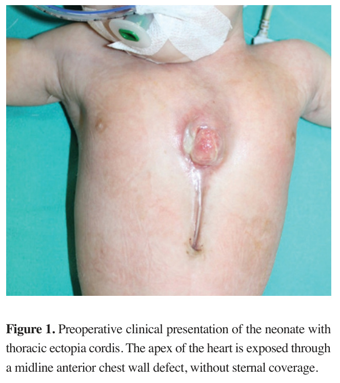
Figure 1: Preoperative clinical presentation of the neonate with thoracic ectopia cordis. The apex of the heart is exposed through a midline anterior chest wall defect, without sternal coverage. |
Echocardiography revealed an atrial septal defect with a left-to-right shunt, a 2 mm patent ductus arteriosus, and an open ductus venosus. Additional findings included aplasia cutis, retrognathia, and a high-arched palate. Thoracic computed tomography (CT) demonstrated complete sternal aplasia with intact bilateral clavicles and ribs, together with a 2 cm anterior thoracic wall defect through which the cardiac apex herniated. Dextrocardia was also noted.
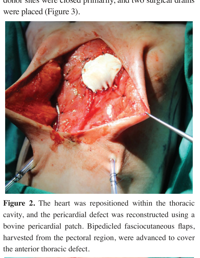
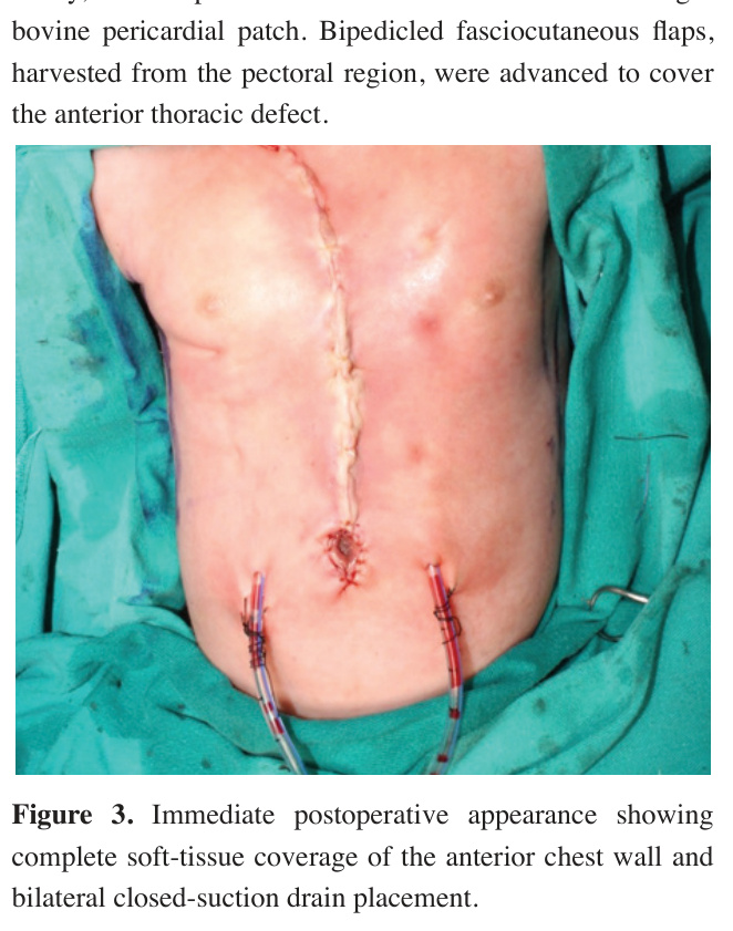
On day 29 of life, after hemodynamic stabilization, completion of diagnostic evaluations, and multidisciplinary operative planning, the patient underwent a combined cardiovascular and plastic surgery procedure. During the preoperative period, the exposed cardiac tissue was managed with saline-soaked dressings to maintain tissue hydration. Antibiotic ointments and advanced wound coverings were intentionally avoided because of concerns regarding possible local reactions and irritation over the exposed cardiac structures. For infection prophylaxis, intravenous ampicillin and gentamicin therapy was initiated and continued throughout the surgical procedures and postoperative period. The cardiovascular surgery team repaired the pericardial defect with a bovine pericardial patch (4 x 6 cm, 0.1-0.5 mm in thickness) and repositioned the heart apex within the thoracic cavity. Cardiopulmonary bypass was not required during the procedure. Simultaneously, chest wall reconstruction was performed using 15 × 15 cm bipedicled fasciocutaneous flaps harvested from the pectoral region (Figure 2). Flap donor sites were closed primarily, and two surgical drains were placed (Figure 3).
)
)
 Click Here to Zoom |
Figure 2: The heart was repositioned within the thoracic cavity, and the pericardial defect was reconstructed using a bovine pericardial patch. Bipedicled fasciocutaneous flaps, harvested from the pectoral region, were advanced to cover the anterior thoracic defect. |
 Click Here to Zoom |
Figure 3: Immediate postoperative appearance showing complete soft-tissue coverage of the anterior chest wall and bilateral closed-suction drain placement. |
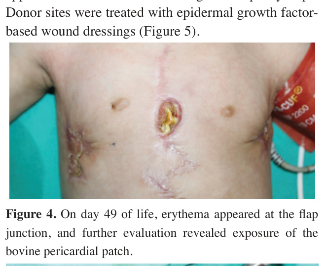
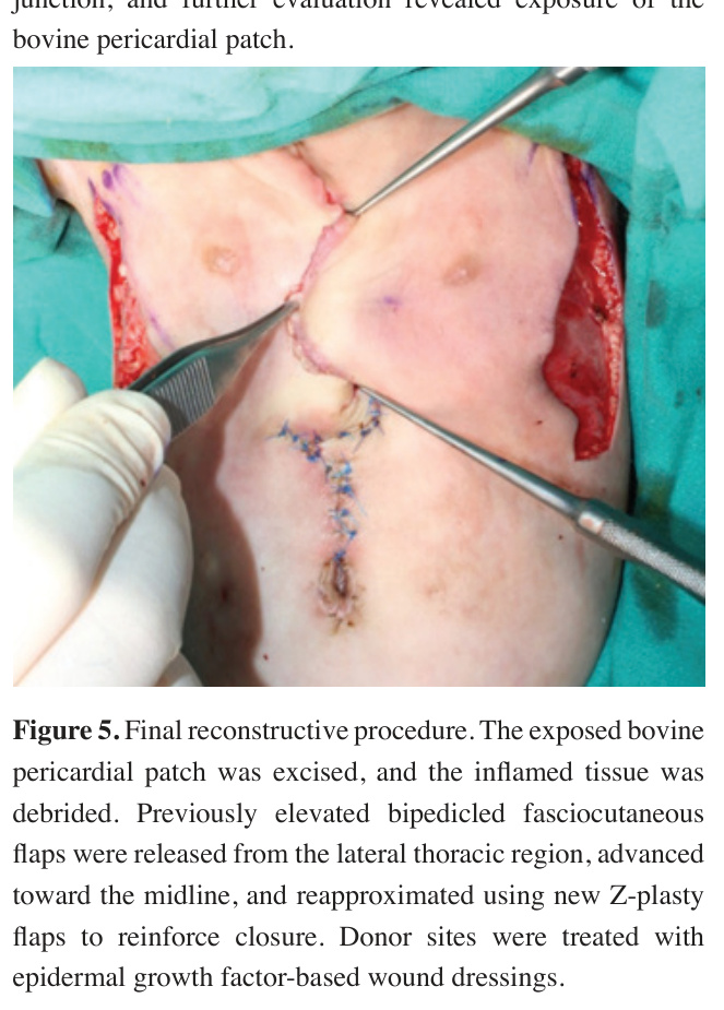
On day 35 of life, necrosis along the inferior suture line was noted, requiring a second surgical intervention. The necrotic tissue was debrided, and Z-plasty was performed for wound closure. On day 49 of life, erythema developed at the flap junction, and further evaluation revealed patch exposure localized to the flap suture line (Figure 4). Wound culture demonstrated no significant bacterial growth. The bovine pericardial patch was excised, and the inflamed area was trimmed. To improve wound closure, previously elevated bipedicled fasciocutaneous flaps were released via lateral thoracic incisions, advanced medially, and approximated at the midline using new Z-plasty flaps. Donor sites were treated with epidermal growth factorbased wound dressings (Figure 5).
)
)
 Click Here to Zoom |
Figure 4: On day 49 of life, erythema appeared at the flap junction, and further evaluation revealed exposure of the bovine pericardial patch. |
 Click Here to Zoom |
Figure 5: Final reconstructive procedure. The exposed bovine pericardial patch was excised, and the inflamed tissue was debrided. Previously elevated bipedicled fasciocutaneous flaps were released from the lateral thoracic region, advanced toward the midline, and reapproximated using new Z-plasty flaps to reinforce closure. Donor sites were treated with epidermal growth factor-based wound dressings. |
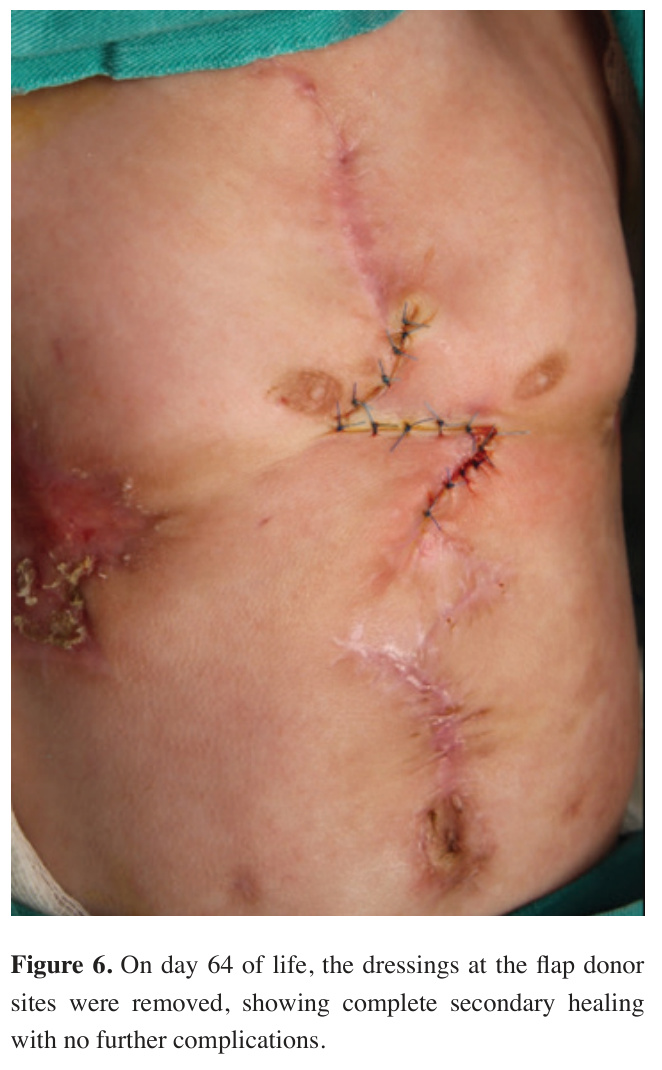
By day 64 of life, the dressings were removed, demonstrating complete secondary wound healing without additional complications (Figure 6). The patient remained hospitalized for an additional 10 days and was discharged in stable condition. Postoperative followup continued for 2 months, during which no recurrent wound complications, signs of infection, hemodynamic instability, or additional cardiac complications were observed. After the second postoperative month, the patient did not return for further follow-up at our institution. To the best of our knowledge, no mortality was reported during the available follow-up period.
)
 Click Here to Zoom |
Figure 6: On day 64 of life, the dressings at the flap donor sites were removed, showing complete secondary healing with no further complications. |
Discussion
Surgical decision-making depends on the type and severity of the defect, associated anomalies, and overall patient stability. Surgical correction remains the definitive treatment [2]. Although successful single-stage repairs have been reported, staged reconstruction may be necessary to allow adequate tissue adaptation and to prevent hemodynamic compromise [1,3,5]. Premature closure of the thoracic cavity may result in compression of the heart and great vessels, necessitating alternative reconstructive strategies such as subcutaneous pockets, tissue expansion, or staged soft tissue advancement [3]. In patients with complete or near-complete sternal aplasia, achieving durable chest wall coverage remains particularly challenging because of limited local tissue availability and increased wound tension. In our patient, postoperative wound complications were likely related to increased tension at the midline closure rather than prosthetic infection, as wound cultures demonstrated no significant bacterial growth.
In the present case, bipedicled fasciocutaneous flaps harvested from the pectoral region were preferred because they provided reliable vascularity, adequate soft tissue coverage, and preservation of thoracic wall compliance without muscle sacrifice. However, postoperative wound complications developed, including suture line necrosis followed by patch exposure. Management of prosthetic exposure in neonatal chest wall reconstruction remains challenging because wound closure and preservation of chest wall stability must be achieved simultaneously. Therefore, a staged reconstructive approach was preferred in our patient. Debridement, excision of the patch, flap release, medial advancement, and repeat Z-plasty reconstruction allowed preservation of chest wall integrity while achieving successful wound closure.
Recent surgical advances have improved the management of ectopia cordis.
Extracorporeal membrane oxygenation (ECMO) has been used in infants developing cardiac failure following repair [3]. Additionally, three-dimensional printing technologies may facilitate preoperative planning and improve chest wall reconstruction accuracy. Alloplastic materials, including synthetic meshes and bioabsorbable plates, may provide structural support and improve long-term reconstructive outcomes. Mohan et al. emphasized the importance of stable soft tissue coverage in pediatric chest wall reconstruction, particularly in patients requiring prosthetic support [4]. In our case, a soft tissue– based reconstructive approach was preferred because it provided adequate chest wall coverage while preserving thoracic compliance during the neonatal period.
Future research should focus on refining surgical techniques, exploring novel biomaterials, and developing individualized reconstructive strategies for these complex patients. Advanced imaging modalities, including fetal MRI, may improve prenatal diagnosis and facilitate earlier multidisciplinary planning. Long-term followup studies remain necessary to better evaluate cardiac function, respiratory mechanics, chest wall development, and quality of life in surviving patients [3]. In selected patients with significant intracardiac anomalies, additional palliative procedures such as Blalock-Taussig shunting or pulmonary artery banding may also be required [6].
Ectopia cordis remains one of the most challenging congenital thoracic anomalies and requires individualized surgical planning. The successful staged reconstruction of our patient with complete sternal aplasia highlights the importance of flexible reconstructive strategies and multidisciplinary management. Continued advances in neonatal care, biomaterials, and reconstructive techniques will be essential for improving survival and long-term outcomes in these patients.
This report has several limitations. The patient was lost to follow-up after two months, precluding assessment of long-term chest wall growth, respiratory mechanics, prosthetic durability, and developmental outcomes. Furthermore, because this is a single case report, conclusions regarding the superiority of this reconstructive strategy cannot be generalized.
In conclusion, ectopia cordis remains a rare and challenging congenital anomaly associated with high morbidity and mortality. Successful management requires individualized multidisciplinary planning involving neonatal intensive care, cardiovascular surgery, and reconstructive surgery teams. In our case, bipedicled fasciocutaneous flaps provided effective chest wall coverage following cardiac repositioning and allowed successful reconstruction after postoperative wound complications. Careful postoperative monitoring and staged reconstructive management are critical for preserving thoracic stability and optimizing clinical outcomes in these complex patients.