
Abstract
Introduction
has considerably expanded the scope of minimally invasive thoracic procedures [1]. Nevertheless, the simultaneous treatment of mediastinal and pulmonary pathologies remains technically challenging because these operations often require different patient positions, port configurations, or re-docking of the robotic system. As a result, combined procedures are frequently associated with increased operative complexity, prolonged anesthesia time, and higher resource utilization.
We present a rare case of a patient with myasthenia gravis (MG) and a synchronous hypermetabolic right middle lobe pulmonary nodule who underwent simultaneous robotic total thymectomy and right middle lobectomy during a single operative session. This approach demonstrates a practical and efficient strategy for multi-compartment thoracic surgery while preserving the ergonomic advantages of the robotic platform and avoiding intraoperative realignment of the system.
The presented case is one of the few reported cases of simultaneous robotic total thymectomy and anatomical pulmonary lobectomy performed through a single docking strategy, without patient repositioning, using a modified periareolar thymectomy port configuration.
Case Presentation
)
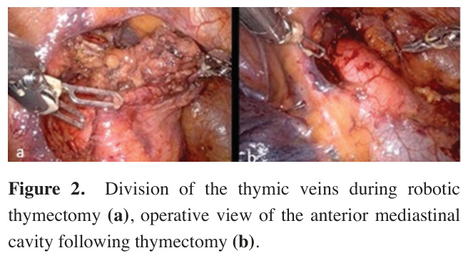
The operation was performed in a near-supine position with minimal table tilt. Initially, robotic total thymectomy was completed using a right-sided periareolar approach with robotic arms positioned through ports 1, 2, and 3 (Figure 2).
)
 Click Here to Zoom |
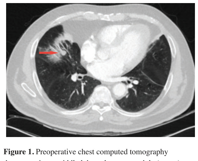
Figure 1: Preoperative chest computed tomography |
demonstrating a middle lobe pulmonary nodule (arrow).
 Click Here to Zoom |
Figure 2: Division of the thymic veins during robotic thymectomy (a), operative view of the anterior mediastinal cavity following thymectomy (b). |
During the thymectomy phase via the right hemithorax approach, the right lung was collapsed with the aid of carbon dioxide insufflation maintained at a pressure of 8-10 cmH2O. The mediastinal pleura was then dissected in a caudal-to-cranial direction, extending up to the junction of the superior vena cava and the brachiocephalic vein. At this stage, gas insufflation facilitates retrosternal and prepericardial dissection by creating a controlled pneumomediastinum. The thymic veins were carefully isolated, divided at their drainage point into the brachiocephalic vein, and secured using titanium clips. Dissection was continued until the left mediastinal pleura was reached, achieving a complete total thymectomy. The resected thymus was placed in an endobag and extracted through the port incision.
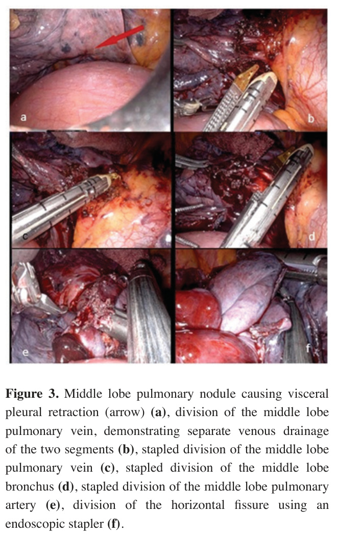
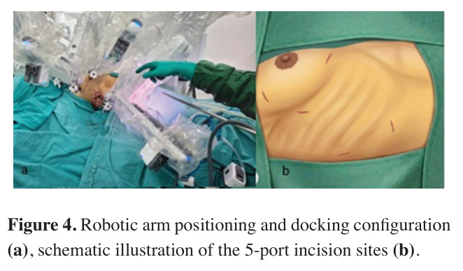
Following completion of the thymectomy, the port configuration was adapted for the pulmonary resection phase without patient repositioning or robotic redocking. A fourth robotic port was added and the robotic setup was reconfigured without re-docking. The lobectomy phase was performed with Arm 4 in port 1, Arm 1 in port 2, the camera arm (Arm 2) in port 3, and Arm 3 in the newly created fourth port (Figure 3). This configuration provided a more favorable working geometry for hilar dissection and pulmonary resection while maintaining the advantages of a single-docking approach. Consequently, the lobectomy was performed using robotic arms 4, 1, 2 and 3, together with a dedicated assistant port (Figure 4).
)
)
 Click Here to Zoom |
Figure 3: Middle lobe pulmonary nodule causing visceral pleural retraction (arrow) (a), division of the middle lobe pulmonary vein, demonstrating separate venous drainage of the two segments (b), stapled division of the middle lobe pulmonary vein (c), stapled division of the middle lobe bronchus (d), stapled division of the middle lobe pulmonary artery (e), division of the horizontal fissure using an endoscopic stapler (f). |
 Click Here to Zoom |
Figure 4: Robotic arm positioning and docking configuration (a), schematic illustration of the 5-port incision sites (b). |
This modification enabled seamless transition from total thymectomy to right middle lobectomy and systematic lymph node dissection while maintaining a single docking strategy throughout the procedure.
During the right middle lobectomy phase, the mediastinal pleura was incised to expose the hilum of the middle lobe under continued carbon dioxide insufflation. The middle lobe veins were observed to drain independently as segmental branches and were subsequently divided through the assistant port using an endoscopic vascular stapler. Immediately proximal to the venous anatomy, the middle lobe bronchus was isolated and divided with a stapler, followed by the dissection and vascular stapler closure of the middle lobe artery. Due to an incomplete horizontal fissure, the resection was completed by creating the fissure line using mechanical staplers. Finally, systematic mediastinal and hilar lymphadenectomy was performed, which included the clearance of stations 4 (lower paratracheal), 10 (hilar), and 12 (lobar) lymph nodes. The resected middle lobe was placed in an endobag and extracted through the assistant port, which had been widened via skin incision extension.
The total operative time was 270 minutes, which included a docking time of 20 minutes and a total robotic console time of 220 minutes. The console phase was divided into 70 minutes for the thymectomy and 150 minutes for the right middle lobectomy. The estimated blood loss was minimal, remaining well below 50 mL throughout the entire procedure.
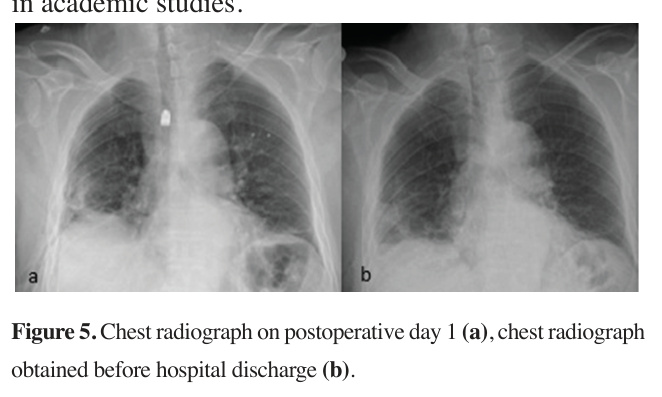
The postoperative course was uneventful. The chest tube was removed on postoperative day four, and the patient was discharged without any complications (Figure
5).
Final histopathological examination revealed invasive adenocarcinoma with 1 cm in greater dimension in the right middle lobe with negative resection margins and no lymph node metastasis (pT1aN0). Histological evaluation of the thymectomy specimen confirmed thymic tissue without evidence of thymoma or malignancy. Written informed consent was obtained from the patient for the use of the medical data in academic studies.
 Click Here to Zoom |
Figure 5: Chest radiograph on postoperative day 1 (a), chest radiograph obtained before hospital discharge (b). |
Discussion
For patients with active MG, this single-stage approach offers a distinct clinical advantage. By consolidating the treatment, it spares the patient from the cumulative risks of repeated general anesthesia and preserves vital postoperative respiratory function by minimizing overall surgical effects on cardiopulmonary function.
Most existing reports on combined resections involve intraoperative repositioning or robotic re-docking, maneuvers that inevitably prolong anesthesia time and disrupt the surgical workflow [2-4].
Okazaki et al described the robotic resection of a posterior mediastinal dumbbell tumor performed with the patient in the prone position [2]. Although such positioning can facilitate surgical exposure, prone position may introduce additional challenges in airway management intraoperatively. This position may also complicate an urgent conversion to open surgery (thoracotomy or sternotomy), as repositioning the patient can be both time-consuming and technically demanding. In contrast, our patient remained in a supine position with a slight lateral tilt throughout the procedure. We believe that this approach not only provided favorable cardiopulmonary conditions for a patient with MG but also allowed the surgical team to convert to the lateral decubitus position or proceed directly to open surgery, if necessary, without repositioning the patient or changing the operating table setup.
The case report reported by Luzzi et al demonstrated that thoughtful robotic planning can simplify complex multi-site procedures [3]. They successfully performed a combined robotic thymectomy and transaxillary thyroidectomy by rotating the robotic platform by only 30° after completing the first procedure, thereby avoiding complete system re-docking. Rather than being merely a technical adjustment, this strategy optimized the use of disposable instruments and contributed to a faster postoperative recovery. Their experience reflects the same principle of our approach. Instead of dismantling and rebuilding the robotic setup between procedures, careful optimization of robot positioning can improve workflow, reduce unnecessary operative steps, and make complex multi-compartment thoracic resections more efficient for both the surgical team and the patient.
Zhang et al further highlighted the technical challenges of multi-compartment robotic surgery by reporting the simultaneous resection of a left-sided anterior mediastinal tumor and a right-sided lower thoracic paravertebral mass through the same four port incisions on the right side of the chest [4]. Although they successfully utilized a single-sided approach with the patient positioned in a left lateral decubitus position throughout, the completely opposite orientations of the two lesions within the right hemithorax required a two-step docking strategy. After completing the posterior paravertebral resection, they had to undock the robotic system, rotate the operating table 180° counterclockwise while keeping the patient in the same decubitus position, and re-dock the system from the ventral side to access the anterior mediastinum. While their technique avoided additional incisions, the need for undocking, table rotation, and re-docking inevitably increases logistical complexity and prolongs both operative and anesthesia times. In contrast, our singledocking strategy for synchronous thymectomy and right middle lobectomy required neither undocking nor operating table manipulation.
In another published case report, simultaneous robotic surgical procedures involving cardiac surgery and lobectomy were documented. The initial approach was performed via a minithoracotomy for the cardiac procedure in the supine position. Following the completion of the cardiac phase, the robotic arms were removed for the lobectomy; the patient was then repositioned into the lateral decubitus position, and robotic docking was established using four ports to perform a right upper lobectomy. The total operative time was reported as 10 hours [5].
In our approach, these limitations were overcome by placing the patient in a supine position with a modest 15-degree right-sided elevation. This configuration provided sufficient exposure for both the anterior mediastinum and the right hemithorax, eliminating the need for repositioning for RATS. The operative sequence was intentionally designed to perform the thymectomy first. This served as a strategic safety measure: by securing the mediastinal clearance while the patient was in the optimal position for thymectomy, we preserved the flexibility to convert to a lateral decubitus position should technical difficulties arise during the lobectomy.
Previously, a simultaneous VATS sleeve lobectomy and thymectomy was described by Xie et al [6]. In that particular case, the surgical team first performed the right upper bronchial sleeve lobectomy in a lateral decubitus position, after which the patient had to be completely repositioned to access the mediastinum for the thymectomy phase. Even within the versatile domain of VATS, where the surgeon typically has a broad range of manual manipulation, a formal intraoperative patient repositioning was still deemed necessary, which stands in sharp contrast to our single-position, single-docking strategy. This highlights a critical reality in multicompartment thoracic surgery: the chosen surgical trajectory and overall approach must adapt dynamically based on which specific pulmonary lobe or mediastinal compartment harbors the pathology.
From an anatomical perspective, the right middle lobe is exceptionally well-suited for this near-supine approach. Its anterior location and favorable hilar orientation allow for a dissection trajectory that closely resembles subxiphoid or anterior thoracoscopic perspectives [7]. Similar to the approach used in the presented case, simultaneous resections may not be appropriate for posterior or superior pulmonary pathologies, such as left-sided resections or lower lobe tumors requiring extensive posterior mobilization, where a standard lateral decubitus position remains mandatory for optimal exposure.
Approaching right-sided pulmonary resections alongside anterior mediastinal surgery through a single incision and a single patient position provides favorable ergonomics and optimal alignment for endoscopic instruments. Indeed, case reports in the literature have demonstrated that simultaneous thymectomy and lobectomy with VATS can be successfully completed via a subxiphoid approach without requiring intraoperative patient repositioning [8]. However, to the best of our knowledge, simultaneous lobectomy and thymectomy performed completely via robot-assisted thoracic surgery (RATS) has not yet been reported.
When evaluating our operative metrics, the docking time in the present case was comparable to existing literature data [9,10]. Although our total console and overall surgical times were longer than those reported for isolated procedures, this discrepancy remains highly acceptable given that our data reflect the cumulative duration of two major resections performed back-to-back.
This technique represents a significant refinement in minimally invasive thoracic surgery. By avoiding the mechanical and logistical delays of re-docking or table manipulation, this single-docking approach maximizes the ergonomic advantages of the robotic platform. For carefully selected patients, this method offers a seamless, efficient, and oncologically sound alternative for concomitant mediastinal and pulmonary resections, ensuring high surgical precision while minimizing perioperative risk.