
Abstract
Introduction
Extraskeletal ES (EES) has an incidence of 0.4/1,000,000 and represents about 20% of ES cases. EES has a bimodal distribution with peak incidence in individuals < 5years and > 35years old. EES, unlike ES of the bone, shows no predilection for biological sex and race. The upper thigh, buttocks, upper arm, and shoulders are the most common sites of EES [2]. The mediastinum is an exceedingly rare primary site of EES and thus presents a diagnostic dilemma due to nonspecific symptoms that may suggest commoner diseases such as Tuberculosis (TB) in high TB-burden countries [3]. We report a presumed case of primary mediastinal ES with a distinctive presentation of pneumothorax and review the literature.
Case Presentation
On examination, she had a temperature of 37.0 0C, blood pressure of 110/70 mmHg, heart rate of 120 beats/ min, respiratory rate of 46 breaths/min, oxygen saturation of 98% in room air, and grade III digital clubbing. Chest examination revealed trachea deviation to the right, stony dull percussion note, and absent breath sounds on the left hemithorax. There was no significant peripheral lymphadenopathy, hepatosplenomegaly, or jaundice.
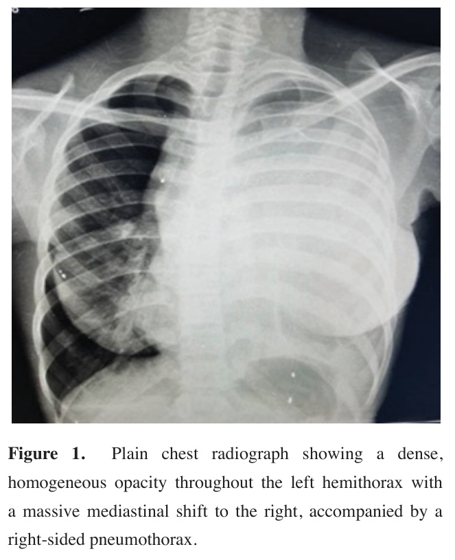
Chest X-ray showed an ill-defined homogenous opacity in the left hemithorax with a mass-induced mediastinal shift to the right, where a pneumothorax was also seen (Figure 1).
)
 Click Here to Zoom |
Figure 1: Plain chest radiograph showing a dense, homogeneous opacity throughout the left hemithorax with a massive mediastinal shift to the right, accompanied by a right-sided pneumothorax. |
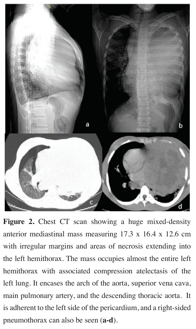
Chest CT scan showed a huge mixed-density anterior mediastinal mass measuring 17.3 x 16.4 x 12.6 cm with irregular margins and areas of necrosis extending into the left hemithorax. The mass occupies almost the entire left hemithorax with associated compression atelectasis of the left lung. It encases the arch of the aorta, superior vena cava, main pulmonary artery, and the descending thoracic aorta. It is adherent to the left side of the pericardium. Right-sided pneumothorax can also be seen (Figures 2a-d). No area of calcification, mediastinal lymphadenopathy, pleural effusion or hepatic metastasis noted.
)
Laboratory evaluation revealed a markedly elevated erythrocyte sedimentation rate (95 mm/hr). Total leucocyte count was 6.3×10⁹/L, packed cell volume of 30%, serum urea: 3.8 mmol/L, creatinine: 75.4 µmol/L, and serum electrolytes were within normal limits. Sputum evaluation was negative for acid-fast bacilli, mycobacterium tuberculosis was not detected on GeneXpert, and cytology was negative for malignancy.
 Click Here to Zoom |
Figure 2: Chest CT scan showing a huge mixed-density anterior mediastinal mass measuring 17.3 x 16.4 x 12.6 cm with irregular margins and areas of necrosis extending into the left hemithorax. The mass occupies almost the entire left hemithorax with associated compression atelectasis of the left lung. It encases the arch of the aorta, superior vena cava, main pulmonary artery, and the descending thoracic aorta. It is adherent to the left side of the pericardium, and a right-sided pneumothorax can also be seen (a-d). |
She had a closed thoracostomy tube drainage for the right-sided pneumothorax and symptomatic treatment. Anti-TB medications were discontinued following the results of laboratory investigations, and she underwent a trans-thoracic percutaneous Tru-cut biopsy of the mediastinal tumor. She was discharged after 7 days of admission following improvement of symptoms and was adequately counseled to see the next scheduled specialist clinic with the histology report.
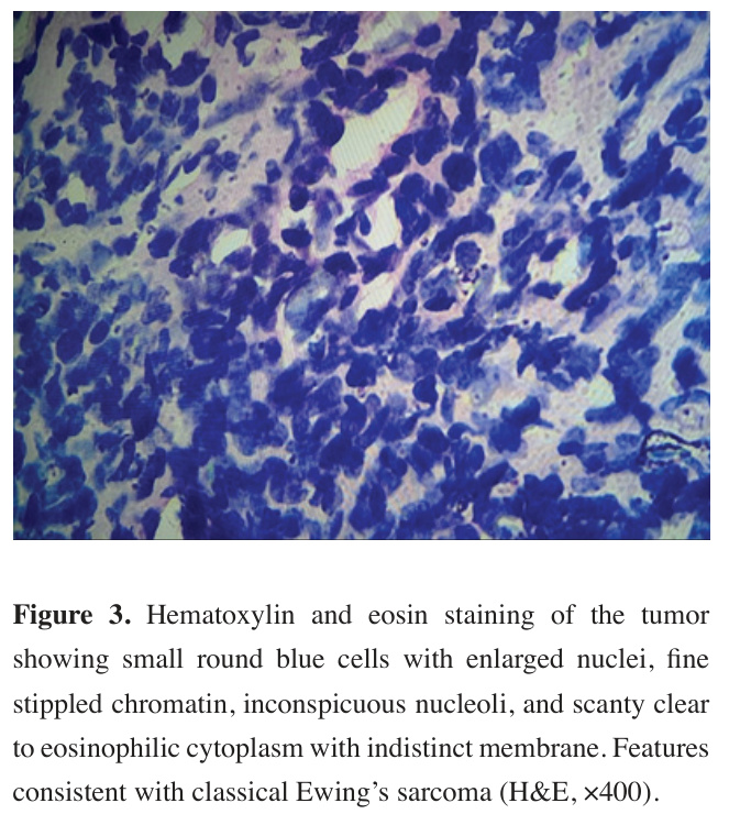
Histological sections showed sheets of monotonous primitive small round blue cells with a high nuclearto-cytoplasmic ratio, hyperchromatic nuclei, some irregular nuclear contours with fine stippled chromatin, inconspicuous nucleoli, and scanty cytoplasm with an indistinct membrane. No mitotic activity or areas of necrosis or hemorrhage were seen; features highly consistent with a small round-cell malignancy, such as Ewing’s sarcoma (Figure 3).
)
 Click Here to Zoom |
Figure 3: Hematoxylin and eosin staining of the tumor showing small round blue cells with enlarged nuclei, fine stippled chromatin, inconspicuous nucleoli, and scanty clear to eosinophilic cytoplasm with indistinct membrane. Features consistent with classical Ewing’s sarcoma (H&E, ×400). |
Unfortunately, our patient only re-presented after four weeks of initial discharge with worsening of her symptoms, as her parent had attributed her illness to a “spiritual attack”, which stems from a magicoreligious thought system of disease causation that is not uncommon in our sub-region [4]. In a sad turn of events, our patient succumbed to her illness within 2 days of re-presentation to the clinic. A deceased patient’s permission form was signed by the patient’s next of kin for the publication of this case report.
Discussion
Imaging plays a vital role in the management of EES. Although CT scan features are non-specific, they remain vital for assessing tumor size and resectability. Commonly seen features include a slightly hypodense soft tissue mass with areas of cystic degeneration on non-contrast CT, while contrast-enhanced medium demonstrates a mass with a mixed-density pattern. Calcification is seen in 10% of cases. Pleural effusion, pneumothorax, and pericardial effusion are manifestations of invasion of nearby structures that may be seen [6-8].
Histopathological analysis is required to confirm the diagnosis of EES. The histological picture of EES consists of a large number of uniform small round cells with a high nuclear-to-cytoplasmic ratio and scanty clear to eosinophilic cytoplasm [9].
Immunohistochemistry (IHC) is an indispensable tool in the diagnosis of ES. Positive staining for Cluster of differentiation 99 (CD99), Friend leukemia integration 1 (FLI-1), and NKX2 are currently acceptable markers for diagnosis of ES on IHC. Additionally, genetic testing is helpful for definitive diagnosis, and the presence of EWSFLI1 fusion gene defines ES on genetic testing [9-11].
What makes this case particularly unique is the rare clinical presentation of a primary mediastinal tumor skewed to the left co-occurring with a contralateral pneumothorax and resultant paradoxical mediastinal deviation.
In classic physiology, a right-sided pneumothorax exerts a positive pressure effect that shifts mediastinal structures toward the left hemithorax. However, in our patient, physical and radiological examinations revealed a severe tracheal and mediastinal shift to the right. This paradoxical finding is explained by the massive volume and dominant compressive force of the left-sided anterior mediastinal mass, which completely overwhelmed the contralateral pressure of the right pneumothorax. Such a presentation can easily mislead clinicians, especially in resource-limited settings where symptoms might be misattributed solely to common infectious pathologies like tuberculosis.
A major limitation of this report is the lack of immunohistochemical (IHC) confirmation and molecular or genetic testing (such as detecting the EWSR1-FLI1 fusion gene), which are vital to establish a definitive diagnosis of Ewing's sarcoma. Due to an inadequate tissue block from the initial biopsy, coupled with the patient's late re-presentation and rapid demise, these ancillary studies could not be completed. Consequently, a broad differential diagnosis for small round blue cell tumors of the mediastinum must be considered. This includes lymphoma, embryonal rhabdomyosarcoma, neuroblastoma, poorly differentiated synovial sarcoma, and newer entities like BCOR or CIC-rearranged sarcomas [12]. While the cytomorphological features were highly suggestive of extraskeletal Ewing's sarcoma, the diagnosis remains presumptive rather than definitive.
Early diagnosis and treatment can improve the prognosis of EES. Treatment involves a combination of systemic chemotherapy and local control either by surgery or radiotherapy. Generally, induction with multidrug chemotherapy is given before local control. Surgery is the preferred method of local control for resectable tumors, as the long-term outcome when radiotherapy is used alone is less favorable, even though ES is radiosensitive [13,14]. Autologous stem cell therapy has also been used in the management of EES [9].
Our literature search identified six documented cases of primary mediastinal ES. Three of the cases were in males and 3 females; the ages ranged from 15 to 66 years. As in our case, the anterior mediastinum was the primary site of the tumor in 3 cases; 2 were located in the posterior mediastinum, and 1 involved both the anterior and posterior mediastinum. Cough was present in 3 out of 4 cases involving the anterior mediastinum, a finding fairly consistent with this index case, while pain was the predominant symptom in the posterior mediastinal tumors. There was an associated pleural effusion in 3 of the 6 cases, and 1 of the 3 cases with pleural effusion had pericardial effusion in addition. Notably, our case is distinguished by the presence of a primary mediastinal small round cell tumor complicated by pneumothorax, a finding we believe is unprecedented in prior case reports. At a minimum, all cases were diagnosed using histology, while 5 included further IHC testing and 3 involved genetic studies. Four of the six patients had surgery as their primary treatment [3,5,7-9,15].
In Table 1, we summarize the management and outcome of six cases of primary mediastinal ES previously reported in the literature.
)
Primary Site Description Year
Sex complaints
Li X et
15/F Cough
Anterior meDiagnosis
15.7x11.7x10.5 cm moderately heterogenous enhancing mass with multiple mediastinal nodes and invading left lung with ipsilateral pleural effuSu C
32/M Chest pain
Posterior
Histology +
4x4x3 cm well-defined mass with heterogenous density
2024
Hale19/F Pain in the
Posterior testing
16x15x15cm heterogenous mass with smooth edges extending to the left hemithorax and supraclavicular region, plus contralateral mediastinal
Cui M
66/M Chest tightAnterior meGiant cystic and solid mass with moderate enhancement and enlarged mediastinal
Invasion of SVC, Pericardium with pericardial effusion, and right lung with ipsilateral pleuCal30/F Cough
Anterior meWell-defined solid mass extending to the left hemithorax compressing the superior lung lobe and contralateral medias2023
Ata F
16/M Cough
Anterior and stem cell therapy.
transplant
Histology
N/A
N/A
18cm mediastinal mass extending to the left hemithorax with ipsilateral pleural effusion and contralateral basal nodule and mediastinal shift
Abbrev.: M: Male, F: Female, CT: Computed tomography, IHC: Immunohistochemistry, N/A: Not applicable (i.e.: details not
In conclusion, primary mediastinal tumors consistent with Ewing’s sarcoma are rare, aggressive malignancies that pose significant diagnostic challenges. This case highlights a highly unusual presentation characterized by a contralateral pneumothorax and paradoxical mediastinal shift. In high-burden regions, such presentations can mask underlying malignancies and lead to empirical tuberculosis treatment, delaying diagnosis. Although the lack of immunohistochemical and molecular confirmation in this case limits a definitive diagnosis, clinicians should maintain a high index of suspicion for small round cell malignancies when evaluating massive mediastinal tumors with atypical intrathoracic pressure dynamics.