
Abstract
Introduction
The narrowest part of the esophagus is the first part at the level of cricopharyngeus, where the cricopharyngeal muscle plays a role to shorten the diameter of the lumen. This anatomical feature led Jackson to name this constricted area the pass of “Bal-el Mandeb” or the "gate of tears" [7].
In this article, we illustrate seven different case scenarios of esophageal perforation resulting from penetrating ingested foreign bodies, a firearm injury, and iatrogenic false intubation. In all seven cases, early surgical intervention provided a safe and effective approach for foreign body removal and complication management, which successfully minimized morbidity, mortality, and the duration of hospitalization.
Case Presentation
Presenting symptoms included throat pain, dysphagia, excessive salivation, gagging, regurgitation, and pleural collection on chest radiographs. Routine posteroanterior (PA) and lateral chest radiographs were obtained on arrival. Multi-slice computed tomography (MSCT) of the chest was ordered after failed endoscopic extraction, when a dangerous complication was suspected, or when pleural effusion was present on initial radiograph.
Postoperative courses varied markedly.
One mortality occurred (secondary to aorto-esophageal fistula); the remaining six patients survived. Table 1 shows a brief summary of the cases.
)
Written informed consent was waived by the Medical Ethics Committee of our institution due to the retrospective nature of the study, use of anonymized data, and minimal risk to patients. However, we informed all participants during the telephone followup and obtained their verbal informed consent.
Cases 1, 2 and 3: impacted disc battery ingestion Case 1
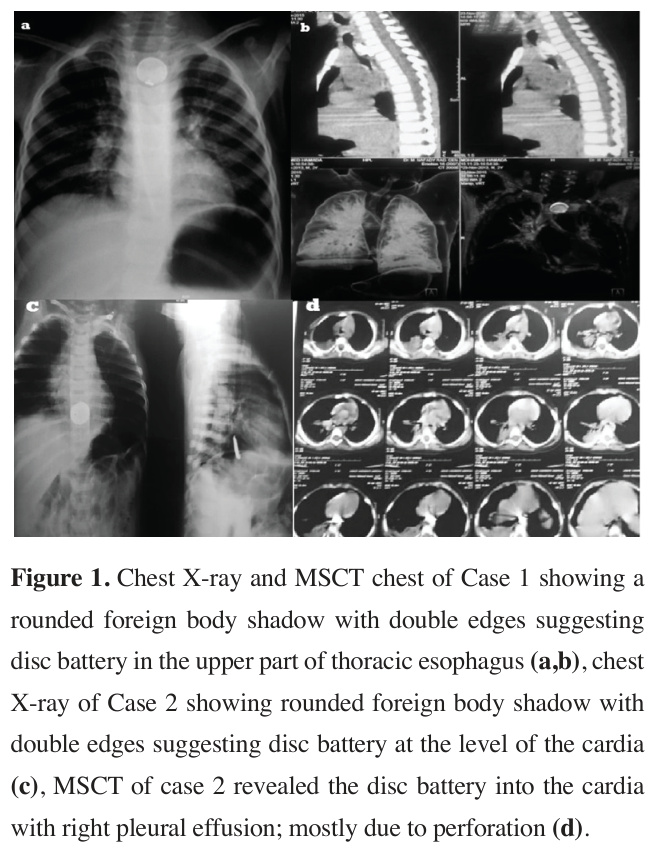
A 15-month-old male child presented with a rounded foreign body impacted in the upper esophagus of unknown nature and duration. A chest radiograph revealed rounded foreign body shadow with double edges suggesting disc battery (Figures 1a,b). The patient was prepared for esophagoscopy which revealed markedly inflamed hyperemic, and easily bleeding mucosa at the suspected site of the foreign body. It was neither possible to extract the foreign body nor to clearly visualize it as it was covered with extensive food particles. Therefore, open surgical extraction was planned.
)
A left half collar cervical incision was performed upon which we found remarkable adhesions around the esophagus. Intraoperative fluoroscopy was used to perfectly locate the foreign body, and a small incision was made in the esophageal wall. The foreign body was confirmed to be a disc battery, that was deeply impacted in and firmly adherent to the esophageal wall. It was covered by granulation tissue and extracted under fluoroscopic guidance. The wound was closed in layers leaving a rubber drain.
Oral feeding was initiated on the fourth postoperative day alongside temporary parenteral nutrition. There was a minimal, transient salivary leak, however, oral feeding was safely continued, the fistula closed spontaneously within a week, and the patient was discharged in good condition on the tenth postoperative day.
Case 2
A 10-month-old female child presented with a history of a disc battery ingestion that occurred more than 10 hours prior while playing with her toys. She was referred to us after failure of multiple esophagoscopic trials of extraction outside our center. A chest radiography and CT was ordered, which revealed distal migration of disc battery to the cardia accompanied by a right pleural effusion, highly indicative of perforation (Figures 1c,d).
Table 1: Summary of complex cases of esophageal perforations. MediastiNo Age/
Etiology
Injury-toDiagnosis-tonitis
Sex
Diagnosis
Surgery
No
> 15 hours Urgent (after
1
Impacted disc failed endosmo/M battery (failed copy)
endoscopy)
Yes
>10 hours
Within 24
2
Impacted disc (right (ingestion hours of mo/F battery (failed pleural to presentareferral (after multiple eneffusion, CT)
doscopies)
tion)
perforation)
No (but >22 hours
Urgent (after
3
2 yr/F Impacted disc esophagoshad battery (with aorto-escopy)
hematemesis)
ophageal fistula)
Acute (<24
Urgent (after
4 yr/M Ingested syNo
4 ringe needle
CT confirmahours from penetrating ingestion)
tion)
aortic arch
Acute (<24
No
Immediately
5
3 yr/M Ingested 10 cm penetrat(after imaghours from ing nail ing)
ingestion)
Firearm inju2 days
Urgent (same
Yes
6 day after CT (diffuse ry to thoracic (food partiyr/M esophagus cles noted diagnosis)
pyogenic in chest memdrain on brane
POD 2)
and food debris) False in>21 days
Urgent (same
Yes
7 tubation (presented day of pres(huge yr/F (iatrogenic,
3 weeks entation)
medidelayed)
post-thyastinal roidectoabscess my)
with barium/ food)
Time to Full
Outcome /
Surgical ApHospital
Oral Intake
Long-term proach
Stay
Follow-up (days)
Survived;
Left cervicoMinimal oral discharged intake at POD tomy + esPOD 10; no
4; fistula ophagotomy + long-term data closed and fluoroscopy reported full intake within 1 week
Right thoracot2 weeks postSurvived; full omy + repair operatively oral intake at (2 tears) +
2 weeks; no gastrostomy long-term data reported
5
Started POD
Died on POD
Rigid es3
5 from massive ophagoscopy + hematemesis extraction secondary to aorto-esophageal fistula
Survived; disLeft posterolat4
48 hours posteral thoracotoperatively charged POD
4; no long-term omy + aortic data reported purse-string
+ endoscopic removal
Right lateral
Survived; dis5
POD 3 charged POD thoracotomy +
5; no long-term primary repair data reported
Right thoraFull at 40
Survived; full days postcotomy + oral intake at primary repair operatively
40 days; no
+ intercostal (slight leaklong-term data muscle flap + age at 2 reported gastrostomy weeks)
Right thoraLight at 1
Survived;
discharged at cotomy + month; full drainage/ at ~2 months
2 months; no decortication (by discharge)
long-term data reported
+ jejunostomy (duodenal perforation)
Abbrev.: POD, postoperative day; M, male; F, female; yr, years; mo, months.
 Click Here to Zoom |
Figure 1: Chest X-ray and MSCT chest of Case 1 showing a rounded foreign body shadow with double edges suggesting disc battery in the upper part of thoracic esophagus (a,b), chest X-ray of Case 2 showing rounded foreign body shadow with double edges suggesting disc battery at the level of the cardia (c), MSCT of case 2 revealed the disc battery into the cardia with right pleural effusion; mostly due to perforation (d). |
The patient was planned for surgical extraction and repair of the esophageal perforation. A right thoracotomy was performed, with ligation of azygous vein for better exploration of thoracic esophagus. The disc battery was extracted, and the tear in distal part of thoracic esophagus was repaired by two layers of interrupted 3/0 polyglactin 910 sutures with meticulous insertion of intercostal tube close to the perforation site. As there was another tear in cervical part of esophagus, the patient was returned to supine position and another cervical collar incision was made and the tear repaired by a single layer of 3/0 polyglactin 910 sutures. Finally, an upper laparotomy incision was made to place a feeding gastrostomy tube for secure enteral nutrition.
Intravenous fluids were initiated postoperatively and gradual enteral feeding via gastrostomy was initiated after 48 hours. Oral fluids commenced one week after surgery with minimal leakage via chest tube, until complete oral intake two weeks postoperatively.
Case 3
A 2-year-old female child presented with repetitive chest infection and hematemesis. Upon examination and radiographic evaluation, there was a rounded impacted foreign body in the lower part of cervical esophagus, mostly disc battery. The patient experienced intermittent, severe attacks of hematemesis, which caused her hemoglobin level to drop sharply from 10.1 g/dL to 6.0 g/dL after each episode.
The patient was planned for rigid esophagoscopy. Under general anesthesia with endotracheal tube in place, the disc battery was successfully extracted with evident slight resistance during extraction which reflects its long-standing impaction into the esophageal wall.
Oral feeding was suspended for 48 hours, after that light fluids were initiated on the third postoperative day. Two days later, the patient developed massive hematemesis and melena. The patient died 2 days later from another massive attack of hematemesis, shock and cardiovascular collapse.
Case 4: ingested syringe needle penetrating the left subclavian artery
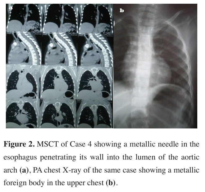
A 4-year-old male child presented with a history of foreign body ingestion (a syringe needle). Chest radiographs were performed, and it revealed a metallic foreign body in the upper chest. The chest radiography and CT revealed a metallic needle in the esophagus penetrating its wall into the lumen of the aortic arch to the left subclavian artery, so the patient was scheduled for open surgical extraction Figures 2a,b).
)
 Click Here to Zoom |
Figure 2: MSCT of Case 4 showing a metallic needle in the esophagus penetrating its wall into the lumen of the aortic arch (a), PA chest X-ray of the same case showing a metallic foreign body in the upper chest (b). |
A limited left posterolateral thoracotomy was performed. The aortic arch and the left subclavian were encircled, a purse string suture was placed around the site of entry of the syringe into the aorta, the foreign body was removed by endoscopy, and the purse string suture was securely tied.
Oral feeding started 48 hours postoperatively, and the patient was discharged on the fourth postoperative day. Case 5: ingested 10 cm nail
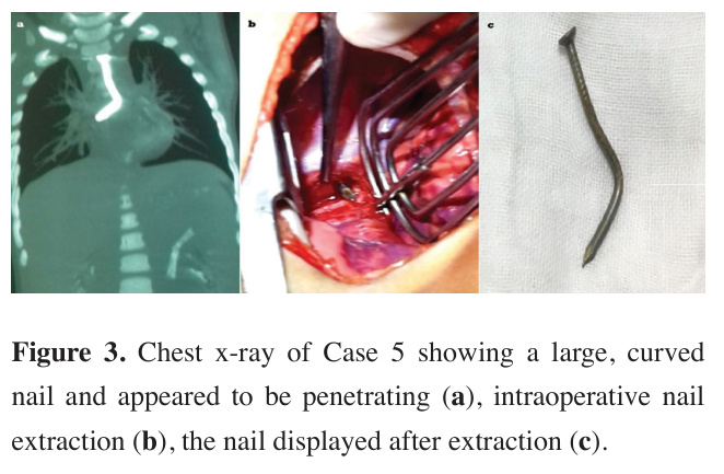
A 3-year-old male child presented with a history of foreign body ingestion (a carpenter's nail). The nail was large and with multiple curvatures and appeared to be penetrating the wall in chest radiograph and CT so open extraction was planned (Figure 3a).
)
A right lateral thoracotomy was performed, and the site of penetration was revealed after opening of the mediastinal pleura and exposure of the thoracic esophagus, and the penetrating nail was visualized and removed (Figures 3b,c). The esophagus was repaired by three simple interrupted 3/0 polyglactin 910 sutures.
Postoperatively, oral feeding was started on the third day and patient was discharged on the fifth postoperative day.
 Click Here to Zoom |
Figure 3: Chest x-ray of Case 5 showing a large, curved nail and appeared to be penetrating (a), intraoperative nail extraction (b), the nail displayed after extraction (c). |
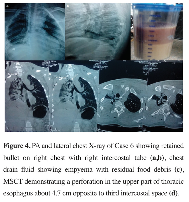
Case 6: firearm injury to the thoracic esophagus A 23-year-old male presented with a gunshot wound to the chest, with an entry wound at the left supraclavicular area and a retained bullet located below the right scapula. On evaluation, he was hemodynamically stable with right hemopneumothorax so right intercostal tube inserted and 500 mL of blood was drained. On the second day after admission, food particles were noticed in the chest drain so chest CT with a water-soluble contrast medium was ordered that revealed perforation in the upper part of thoracic esophagus about 4.7 cm opposite to the third intercostal space. The patient was planned for urgent surgery (Figures 4a-d).
)
 Click Here to Zoom |
Figure 4: PA and lateral chest X-ray of Case 6 showing retained bullet on right chest with right intercostal tube (a,b), chest drain fluid showing empyema with residual food debris (c), MSCT demonstrating a perforation in the upper part of thoracic esophagus about 4.7 cm opposite to third intercostal space (d). |
A right lateral periscapular thoracotomy incision was performed, and the chest was entered through the fourth intercostal space. Before entering the chest, the bullet was extracted below the inferior angle of right scapula. After opening the chest (pleural cavity), intraoperative evaluation revealed a diffuse pyogenic membrane covering the right lung and the mediastinum with food debris in the right thoracic cavity, and about 5 cm esophageal tear. The tear was repaired by 3/0 polyglactin 910 sutures reinforced by vascularized intercostal muscle flap, and the mediastinum irrigated with saline. A wide bore right chest drain was placed, and thoracotomy was closed in layers in a standard fashion. The patient then placed in supine position and mini laparotomy incision was made for placement of feeding gastrostomy.
Postoperatively, nothing per mouth and parenteral nutrition were started immediately. Feeding through gastrostomy tube started on the third postoperative day. Two weeks later, the patient was allowed to begin slight oral intake of light fluids increasing gradually over the days and that associated with slight leakage from chest tube. The patient allowed full oral intake after 40 days postoperatively, and the subsequent course was uneventful. Case 7: sharp esophageal injury due to false intubation
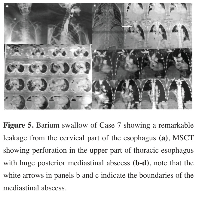
A 43-year-old female patient was referred to our department in septic shock, with a history of a total thyroidectomy performed 3 weeks prior. On evaluation, she was hemodynamically unstable with septic shock and tender rigid abdomen, sweaty, tachycardia and hypotension which required infusion of 60 ng/kg/min of norepinephrine. Additionally, there was a right chest drain showing food particles and empyema. Radiological evaluation showed a large tear in the cervical part of the esophagus with remarkable leakage, as well as huge cervical abscess extending into the superior mediastinum. Barium swallow and chest MSCT demonstrated massive leakage from the proximal cervical esophagus into the superior posterior mediastinum (Figures 5a-d) and her laboratory studies revealed WBCs count: 65,000/µL, together with hypoproteinemia and impairment of renal function. Urgent exploration of the esophagus via right thoracotomy incision was indicated.
)
 Click Here to Zoom |
Figure 5: Barium swallow of Case 7 showing a remarkable leakage from the cervical part of the esophagus (a), MSCT showing perforation in the upper part of thoracic esophagus with huge posterior mediastinal abscess (b-d), note that the white arrows in panels b and c indicate the boundaries of the mediastinal abscess. |
Intraoperative evaluation revealed there was a huge mediastinal abscess with contamination with food particles and barium contrast material. Mediastinal drainage and irrigation were done together with decortication of the right lung. Two wide-bore right chest drains were placed, and thoracotomy was closed in layers in a standard fashion. The patient was then placed in supine position, and upper mini laparotomy incision was made for placement of feeding gastrostomy, but surprisingly we found large perforated duodenal ulcer (stress ulcer) which was repaired by omental patch (Graham's patch), in addition to peritonitis so feeding jejunostomy was the choice and abdominal drains were placed.
Postoperative course of the patient was remarkably slow due to the long-standing perforation more than 21 days and the subsequent toxemia. Parenteral nutrition was initiated immediately postoperatively together with feeding through jejunostomy tube. Significant leakage from cervical tear into thoracic drains continued postoperatively which started to decline 3 weeks postoperatively. Light oral fluids started one month after surgery. Leakage around jejunostomy tube added further impedance to the postoperative course. Upper endoscopy was performed to assess the perforation and possibility of stenting of upper esophageal tear. Upper endoscopy revealed inability to place a stent as the tear was very proximal. The duodenal ulcer was found to be completely healed. Continuation of feeding via jejunostomy and nasogastric tube continued. After 2 months postoperatively, the patient started light oral fluids and the patient was discharged.
Discussion
Most foreign bodies (80-90%) will pass spontaneously through the normal pathway of gastrointestinal tract in about one week without causing any noticed complications; however, 10-20% will need endoscopic removal and nearly 1% will need surgery [8].
Foreign body impaction usually presents acutely with a clear history of ingestion especially for adult patients who might have diffuse chest pain, heaviness, sensation of nausea, vomiting or even choking, sometimes vague foreign body sensation, and neck or throat pain. On the other hand, children may have vague history of refusal of feeding, crying or even vomiting [8].
Removal of a foreign body from the esophagus is mandatory, as its impaction leads to mucosal ulceration, inflammation and subsequently morbid complication such as lung abscess, empyema, mediastinitis, perforation and even a fistula between aorta and esophagus [9]. If endoscopic extraction fails, surgery is the treatment of choice. Different surgical approaches can be performed according to the location of the foreign body which might be cervicotomy, thoracotomy or gastrostomy [9].
Contemporary non-operative and minimally invasive alternatives
In recent years, the management of esophageal perforations has evolved significantly with the introduction of endoscopic and minimally invasive techniques. Covered self-expanding metal stents (cSEMS) have emerged as an effective treatment option, particularly for iatrogenic perforations and contained leaks, with a pooled sealing rate of 86.1% (95% CI: 80.2–92.0%) and stent-related mortality of 7.4% (95% CI: 3.5-11.4%) reported in a recent metaanalysis [10]. Endoscopic vacuum therapy (EVT) is another promising organ-preserving approach, based on negative pressure wound therapy, with reported success rates of 89% in multicenter cohorts [11]. The VACStent, a novel device combining a fully covered stent with an integrated EVT sponge has shown an 80% leak healing rate with no mortality in early multicenter studies [12]. Both esophageal stenting and EVT show high efficacy in sealing esophageal defects, with stenting achieving 86.1% success and EVT demonstrating 89.6% success in sensitivity analyses [10]. These advancements have expanded the therapeutic armamentarium, allowing non-operative management (NOM) to be offered to stable patients with early presentation, contained esophageal disruption, and minimal contamination of surrounding spaces [13].
Rationale for open surgical approach in our series Despite these advances, open surgery remains the cornerstone of management for specific clinical scenarios [13]. In our series of seven cases, operative intervention was chosen based on patient-specific factors that precluded endoscopic or minimally invasive alternatives:
In our case series, esophageal trauma resulted from various etiologies; disc battery ingestion, sharp foreign body, firearm injury and false intubation.
For cases 1and 2, esophageal injury resulted from neglected disc battery ingestion which led to caustic effect and required surgical intervention to remove the disc battery which was deeply and firmly impacted in esophageal wall and to repair the tears resulting from esophagoscopic manipulations.
Endoscopic stenting or EVT would not have addressed the need for foreign body removal, and the presence of extensive local inflammation made these modalities unsuitable. Furthermore, disc batteries in the esophagus are an indication for emergent surgery when they are irretrievable or close to vital structures [13].
In both cases, surgery provided; a) safe extraction of foreign body, b) primary repair of esophageal perforation, c) effective drainage, and d) placement of a port for enteral feeding as in 2nd case.
In case number 3 and 4, we have 2 different types of esophageal trauma and complication. In case number 3, there was neglected disc battery ingestion, presenting with hematemesis, which should have raised suspicion about aorto-esophageal fistula according to previous records [14,15]. But, unfortunately in our situation that was overlooked, and the battery extracted which was followed later on by massive hematemesis and hemodynamic collapse on the 2nd postoperative day. A similar case resulting in patient death due to an aorto-esophageal fistula was also reported in the recent literature [16,17].
Rarely, the diagnosis of aorto-esophageal fistula is known before massive hematemesis occurred. Chiari’s triad of chest pain and sentinel hematemesis followed by rapidly massive hematemesis at a variable interval of time, which is a characteristic feature of aortoesophageal fistula that mentioned in the literature [18], is rarely observed in fistulas resulting from impacted foreign bodies. Sudden death from an aorto-esophageal fistula due to an impacted ingested foreign body is also reported in the literature [16,19].
In our second case of aorto-esophageal fistula (case number 4), the lesson was learnt and as chest CT demonstrated penetration of the needle into the aortic arch, left posterolateral thoracotomy was performed and we controlled the perforation of the aortic arch before removal of foreign body. Endoscopic removal or stenting would have been dangerous, as manipulation could dislodge the needle and precipitate exsanguination.
The above terrible cases of esophageal perforation from foreign body ingestion suggest a public health problem in the region with improvement for intervention to prevent.
The thoracic surgeons should have low threshold to demand chest CT for every case of foreign body impaction in the esophagus even if it looks simple, because it may reveal a complication that not be evident in plain chest radiograph only. Using MSCT chest facilitate shorten the study time and add accuracy to diagnosis as it is considered a useful diagnostic tool with high sensitivity for esophageal foreign body [20,21].
Penetrating trauma to esophagus is uncommon mostly due to its protected position even in busy urban trauma center [22-25]. However, perforation from instrumentation is the commonest such as those occurring during esophagoscopy, and esophageal dilation, frequent use of upper endoscopy, and gastroscopy. The commonest area of perforation occurring during an endoscopic examination is at the level of the cricopharyngeus [26].
Traumatic endotracheal intubation can be another cause of esophageal perforation which occurred just below the cricopharyngeus muscle, generally in the posterior midline at Lannier's triangle (the weakest area where the mucosa is supported only by fascia) [27], and that is what happened in one of our cases.
Selective or conservative management versus mandatory exploration or surgical intervention is still a point of debate in case of penetrating trauma to esophagus [24,28,29].
In all our cases, surgery was the mainstay and immediate management of choice. Because, first, early surgical intervention provides a safe and effective management of cases of esophageal perforation as it provides a tool for adequate mediastinal drainage, irrigation and thus relief of toxemia. Second, surgery allows accurate and meticulous placement of chest tube to drain the perforation site. Third, primary repair of esophageal tear which is effective in early hours of perforation preventing the further complication to occur such as mediastinitis, lung abscess, and empyema which is fatal if not urgently managed. Fourth, in delayed esophageal perforations, repair of tears is very debatable, for some surgeons, they see it ineffective due to edematous edges, inflamed tissues and inflamed mediastinum [30]. For us, we adopt repair for every case of esophageal perforation even delayed perforation either by direct suturing alone or plus augmentation by vascularized tissue. In our opinion, it is associated with decrease fistula size and hence its output and shorter recovery period which coincides with the findings of Wang et al [31]. Lastly, it provides a port for enteral feeding such as gastrostomy or jejunostomy to improve healing.
Surgery in delayed esophageal perforations doesn’t necessarily mean repair of esophageal tear. In our serial cases we performed esophageal repair in late perforations in two cases; that of firearm injury to the esophagus and that of impacted disc battery. Non operative management was not selected, as it associated with failure and high mortality especially if the perforation associated with pleural effusion, mediastinitis and contrast extravasation on chest CT. This coincides with opinion of Merchea et al and Mavroudis et al [32,33].
Postoperative care and surgical nutrition are backbone for success of surgery. In contrast to some authors [23,28], who prefer use of total parenteral nutrition (TPN), we adopt the use of enteral nutrition through gastrostomy tube and that was the opinion of Madiba et al and Muckart et al [24] not only to improve healing, but also to prevent complication from long use of total parenteral nutrition [25,34].
Limitations of the study
Systematic, long-term follow-up beyond hospital discharge was not available for all patients. Outcome data are therefore limited to the immediate postoperative period and, where reported, time to full oral intake. Late complications such as esophageal stricture, dysphagia, or recurrent fistula cannot be excluded.
In conclusion, esophageal perforation remains an exceptionally challenging surgical emergency carrying high risks of morbidity. In this retrospective series of seven patients with diverse etiologies, early surgical intervention achieved survival in six patients. Delayed perforation and aorto-esophageal fistula carry high mortality. Surgery remains the cornerstone of management and was associated with favorable outcomes in most patients, with three key principles: drainage, primary repair (when feasible), and establishment of enteral feeding access. Finally, these cases underscore a critical public health issue in our region, highlighting the urgent need for enhanced parental awareness and regulatory actions to prevent accidental disc battery ingestions in children.