
Abstract
Introduction
Case Presentation
)
 Click Here to Zoom |
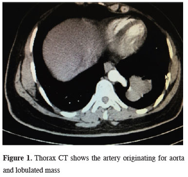
Figure 1: Thorax CT shows the artery originating for aorta and lobulated mass |
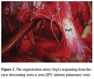
On physical examination, no pathology was detected. The patient was hospitalized with the pre-diagnosis of intrapulmonary sequestration. Fiber-optic bronchoscopy was performed to display the bronchial tree in the preoperative period. Neither any endobronchial lesion nor abnormal bronchial connection that may be associated with the lesion in the left lower lobe was found. A utility incision of 3.5 cm was made through the 6th intercostal space and a 1.5 cm thoracoport incision was placed at the 8th intercostal space and videothoracoscope was placed in the left hemithorax. Adhesions between the left lower lobe and diaphragm were carefully separated. Pulmonary ligament was dissected carefully. The sequestration artery originating from thoracic descending aorta was revealed (Figure 2) and divided with endoscopic vascular stapler. Then the lower lobectomy was completed and did not encounter any complications. A chest drain was placed in the thoracic cavity. The patient did not have any problems during postoperative period and was discharged on the 3rd day following the operation. The pathological result was reported as intralobar sequestration. The patient is still under outpatient clinic follow-up.
)
 Click Here to Zoom |
Figure 2: The sequestration artery (SqA) originating from thoracic descending aorta is seen (IPV; inferior pulmonary vein). |
Discussion
In intralobar sequestration, abnormality is seen as an intrapulmonary lesion in the posterobasal segment of the left lower lobe as displayed in our case. The right lower lobe can be affected in about one third of the cases [2]. Although reports of conservative treatments of pulmonary sequestration, such as spontaneous involution and arterial embolization, have been published, the treatment is commonly surgery even in asymptomatic patients. Before the surgery, antibiotic treatment and respiratory physiotherapy should be performed and if any, infection should be kept under control.
It is proved that VATS is a safe and feasible surgical procedure in experienced hands [4]. The difficulty of resection is to identify the abnormal artery. In the region of sequestration due to frequent recurrent infections, there are usually inflammatory changes and the artery is hidden under the inflamed tissue. There may be very dense adhesions and inflamed tissue may mimic the artery. Abnormal arteries or veins should be identified carefully at the beginning of the procedure [5]. There were adhesions between the lower lobe and diaphragm in our case as well; however, due to the recognition of the intrapulmonary sequestration in preoperative period and display of the abnormal systemic artery, the systemic artery was dissected carefully and divided properly.
As a conclusion, the treatment of pulmonary sequestration is surgery. In such cases, VATS can be safely performed in experienced hands. It is important to perform dissection carefully and to display the abnormal artery. Resection by VATS reduces morbidity and shortens duration of stay in the hospital.
Declaration of conflicting interests
The authors declared no conflicts of interest with respect to the authorship and/or publication of this article.
Funding
The authors received no financial support.