
Abstract
Introduction
Case Presentation
)
 Click Here to Zoom |
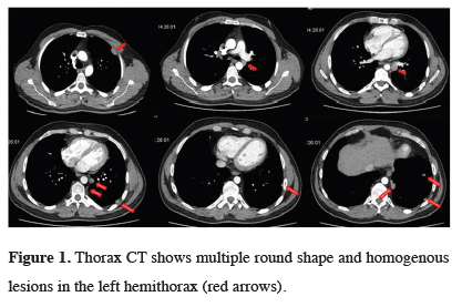
Figure 1: Thorax CT shows multiple round shape and homogenous lesions in the left hemithorax (red arrows). |
Transthoracic fine needle aspiration biopsy was not diagnostic. We decided to perform an exploratory video-assisted thoracoscopic surgery (VATS) for the diagnosis and treatment. However, it was converted to a thoracotomy due to excessive adhesions. Upon exploration, multiple lesions on the chest wall, aortic adventitia, and lung parenchyma were observed. Excisional biopsy of one of the lesions performed and the frozen section reported a collection of lymphoid cells in the vasculature pattern and benign/malign discrimination could not be done on histopathological examination. All masses was resected from the defined regions report.
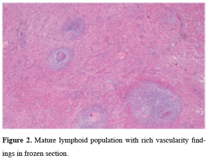
The final pathology examination of the specimens revealed that, there was perivascular mature lymphocyte population that formed nodules in the roof with rich interstitial vascularity which was compatible with typical spleen morphology (Figure 2). When the patient’s anamnesis was detailed, a history of blunt trauma to the abdominal region, which was medically treated 4 years ago, was found.
)
 Click Here to Zoom |
Figure 2: Mature lymphoid population with rich vascularity findings in frozen section. |
Discussion
Shaw et al [2] reported for the first time in 1937 that splenic tissue autotransplantation after splenectomy to the thoracic cavity resulted in an incidence of TS. In 2016, Tulinský et al [3] stated that there were 75 patients in the English literature. A total of 81 cases were published until now. Most of the patients had abdominal trauma in their history. The splenic nodules may be few or up to hundreds in the peritoneal cavity and can be localized in the splenic bed, omentum, mesentery, liver, or diaphragm [4]. Splenic tissue could be found in intraperitoneal or extraperitoneal locations. Splenosis has also been reported rarely in the pericardium, subcutaneous tissue and even in the occipital pole of the brain [1].
The time interval between abdominal injury requiring splenectomy and thoracic splenosis detection varies from several years to decades [3]. Most patients with splenosis are asymptomatic, but 10-15% of them may have pleuritic pain, pleurisy, recurrent hemothorax, or fever [3]. Patients with a diaphragm, phrenic nerve, and parietal pleural involvement may have pain. Our patient was symptomatic because of the location of lesions.
The differential diagnosis of intrathoracic splenosis includes lung carcinomas, pleural metastases, lymphoma, malignant mesothelioma, invasive thymoma, gossypiboma and fibrous tumors of the pleura. Scintigraphy with Tc-99m-labeled and heat-altered erythrocyte is the gold standard method [5,6]. Thorax CT shows a well-defined, homogeneous, pleural-based nodule in the left hemithorax [7,8]. Thorax CT of our patient had multinodular, well-defined masses in the lung. It is not necessary to surgically treat patients who are suspected of TS in the preoperative period.
A 99 m technetium‐labeled heat‐damaged red cell scan is highly specific for detection of splenosis and considered as the gold standard, due to reduced uptake in the normal liver. With the emergence of improved resolution of planar/single photon emission computed tomography/computed tomography (SPECT/CT) scans, Tc‐99 m sulphur colloid scintigraphy can be a reliable for differentiating splenic tissue from non‐splenic tissue [7,9].
Especially in splenectomy patients, surgical resection is unnecessary. Surgical resection should be considered only in symptomatic patients. In our 6 months follow-up after discharge, the patient’s pain was ceased. If there are difficulties in diagnosing on left lower zone lesions, the history of trauma and splenectomy should be questioned in carefully.
Declaration of conflicting interests
The authors declared no conflicts of interest with respect to the authorship and/or publication of this article.
Funding
The authors received no financial support.